Biology 2
Frog
Dissection
Background: The American Bullfrog (Lithobates catesbeianus or Rana catesbeiana)
Frog Taxonomy
| Kingdom | Animalia |
| Phylum | Chordata |
| Class | Amphibia |
| Order | Anura |
| Family | Ranidae |
| Genus | Lithobates |
| Species | catesbeianus |
As members of the class Amphibia,
frogs may live some of their adult lives on land, but they must return to water
to reproduce. Being the first land chordates, they needed to develop or
evolve different characteristics than those of the chordates that lived in the
water. Here are some of those adaptations and how they were an advantage
to terrestrial dwelling chordates:
1. legs: enabled the amphibian to move about the land much easier than the lobe-finned fish ancestors.
2. strong skeleton made of bone: without the water to provide some buoyancy, the amphibian needed a stronger frame to hold up its body while on land.
3. Lungs: lungs are necessary for living on land to extract oxygen from the air. The amphibian lungs are not very efficient so they also use their moist skin as an oxygen exchanger while swimming in water.
4. Nictitating membrane: this is a thin, transparent membrane that covers the eyes. It protects the eyes from debri in the water, but more importantly protects the eyes from drying out while on land.
Here are some other features of the amphibians:
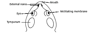
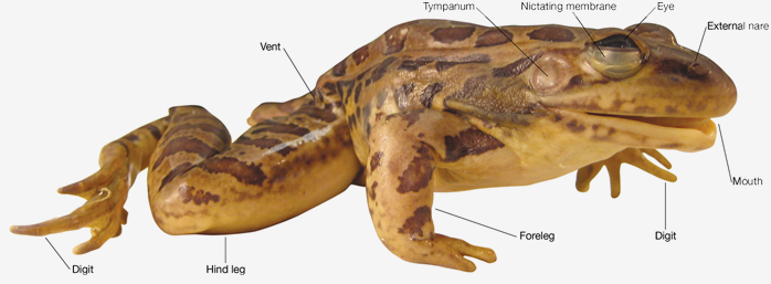
Eggs are laid and fertilized in water. On the outside of the frog’s head are two external nares, or nostrils; two tympanic membranes, or eardrums, which the frog uses to hear with; and two eyes, each of which has three lids. The third lid, called the nictitating membrane, is transparent, it is used to protect the eye from debri and prevent the eye from drying out when on land. Unlike the fish, the placement of the external nares and eyes are on the top of the head instead of the sides and front of the head like the fish. This helps the frog keep its body in the water, while still being able to keep its eyes and nares above the water line. This advantage helps the frog to be able look for predators as well as keep an eye out for food all while being mostly submerged.
You will notice that one of the most obvious features of the frog are its back legs. These legs and feet are well adapted for living both in the water and on land. The strong bone structure and muscular legs are an advantage on land as they can use them to jump quickly away from predators or for catching their own prey. You will also notice that the feet are webbed (webbed toes). This helps the frog to be an efficient swimmer while in the water.
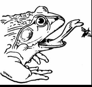
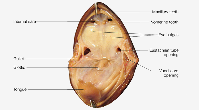
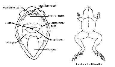
Inside the mouth are two internal nares, or openings into the nostrils; two vomerine teeth in the middle of the roof of the mouth; and two maxillary teeth at the sides of the mouth. These "teeth" are designed to hold their prey while they swallow it whole. They are not intended for chewing. Also inside the mouth behind the tongue is the pharynx, or throat. The tongue of the frog is a bit unusual. Instead of being attached at the rear, the frog tongue is attached at the front. This allows the frog to extend the tongue and retrieve food. See the animated photo below and watch how the tongue moves during the eating phase. The tongue of your specimen, however, is not as pliable. This is due to the fact that it is a muscle and that in the "dead" state of your frog, it is rather stiff.
If you are observant, you will notice that the ventral or belly side of the frog is lighter in color than the top or dorsal side. This coloring scheme is known as counter-shading. Counter-shading allows for camouflage from both above and below viewpoints. From above, the dark colors blend in with the surrounding environment. From below, the light coloring blends in with the light from the sun above. This type of coloration is used by many aquatic animals, as it is quite effective in allowing the animal to blend in.
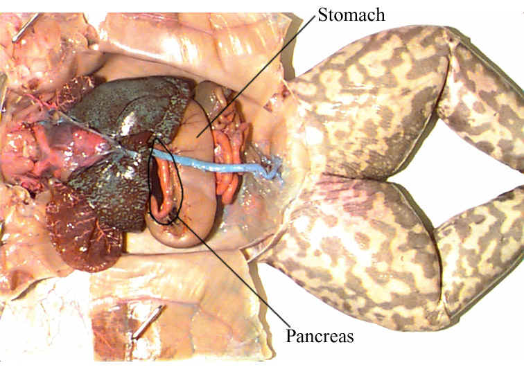
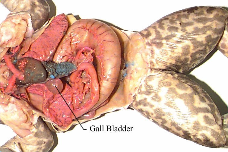
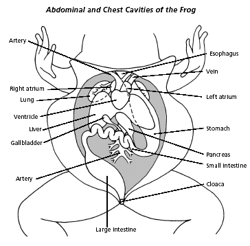
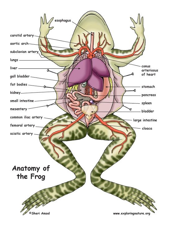
In the pharynx, there are several openings: one is the glottis which opens up into the esophagus, the tube into which food is swallowed; two the larynx, or voice box, air will travel over the larynx and the frog is able to make vocalizations; and three into the Eustachian tubes, which connect the pharynx to the tympanum or tympanic membrane. The digestive system consists of the organs of the digestive tract, or food tube, and the digestive glands. From the esophagus, swallowed food moves into the stomach (stores and churns the food and begins chemical digestion) and then into the small intestine (main organ of digestion, this is where the nutrients are absorbed into the blood stream). Bile is a digestive juice made by the liver (also involved in detoxifying blood and producing proteins needed for digestion) and stored in the gallbladder. Bile flows into a tube called the common bile duct, into which pancreatic juice, a digestive juice from the pancreas, also flows. The contents of the common bile duct flow into the small intestine, where most of the digestion and absorption of food into the bloodstream takes place.
Indigestible materials pass through the large intestine (water is reabsorbed from the undigested food in the large intestine) and then into the cloaca, the common exit chamber of the digestive, excretory, and reproductive systems. The respiratory system consists of the nostrils and the larynx, which opens into two lungs, hollow sacs with thin walls. The walls of the lungs are filled with capillaries, which are microscopic blood vessels through which materials pass into and out of the blood. The circulatory system consists of the heart, blood vessels, and blood. The heart has two receiving chambers, or atria, and one sending chamber, or ventricle. Blood is carried to the heart in vessels called veins. Veins from different parts of the body enter the right and left atria. Blood from both atria goes into the ventricle and then is pumped into the arteries, which are blood vessels that carry blood away from the heart. The spleen is the organ that serves as a reservoir for blood and it also functions to remove old, worn-out blood cells from the circulatory system.
The urinary system consists of the frog’s kidneys, ureters, bladder, and cloaca. The kidneys are organs that filter the blood of impurities. Connected to each kidney is a ureter, a tube through which urine passes into the urinary bladder, a sac that stores urine until it passes out of the body through the cloaca. The organs of the male reproductive system are the testes, sperm ducts, and cloaca. Those of the female system are the ovaries, oviducts, uteri, and cloaca. The testes produce sperm, or male sex cells, which move through sperm ducts, tubes that carry sperm into the cloaca, from which the sperm move outside the body. The ovaries produce eggs, or female sex cells, which move through oviducts into the uteri, then through the cloaca outside the body.
The central nervous system of the frog consists of the brain, which is enclosed in the skull, and the spinal cord, which is enclosed in the backbone. Spinal Nerves branch out from the spinal cord and can be visible in the abdominal cavity of the frog if you look carefully. The frog’s skeletal and muscular systems consist of its framework of bones and joints, to which nearly all the voluntary muscles of the body are attached. Voluntary muscles, which are those over which the frog has control, occur in pairs of flexors and extensors. When a flexor of a leg or other body part contracts, that part is bent. When the extensor of that body part contracts, the part straightens. It is the nervous system that controls many of the frogs senses and behaviors. Here are a few of the structures involved:
Cerebrum: main area of the brain that helps the frog respond to its environment.
Cerebellum: portion of the brain involved with muscle coordination and allows the frog to maintain balance.
Pineal body or pineal gland: small gland in the brain that is involved with the processes of hibernation or estivation.
Optic lobe: portion of the brain devoted to the sense of vision
Olfactory lobe: portion of the brain devoted to the sense of smell
Objectives:
• Describe
the appearance of various organs found in the frog.
• Name the organs that make up various systems of the
frog.
Purpose:
In this lab, you will dissect
a frog in order to observe the external and internal structures of frog anatomy.
Frog dissection has been a staple of biology classes for many, many years.
The reason for this includes:
Internal organs of the frog are in roughly the same location as a human.
Internal organ functions are relatively the same as the human counterparts.
Frogs are relatively inexpensive to raise for the sole purpose of dissections.
Materials:
• forceps
• preserved frog
• dissecting pins (6–10)
• dissecting tray and paper towels
• plastic zip-lock storage bag
• scissors
• marking pen
• dissecting probe
Click HERE for a packet of frog diagrams
Procedure: Video: Frog Dissection External Anatomy
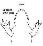
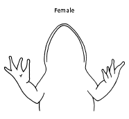
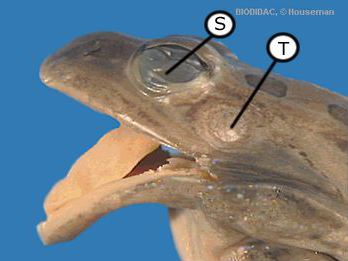
Place a frog on a dissection tray. To determine the frog’s sex, look at the hand digits, or fingers, on its forelegs. A male frog usually has thick pads on its "thumbs," which is one external difference between the sexes, as shown in the diagram below. Another method is to look at the tympanic membrane. Female frogs have tympanic membranes that are approximately the same size as their eye. Male frogs usually have a tympanic membrane that is much larger than their eye.
|
|
|
|
|
|
|
The picture above shows the comparison between the eye and the tympanic membrane. Based on the sizes, this specimen would be a female frog. |
|
Use the diagrams below to locate and identify the external features of the head. Find the mouth, external nares, tympani, eyes, and nictitating membranes.
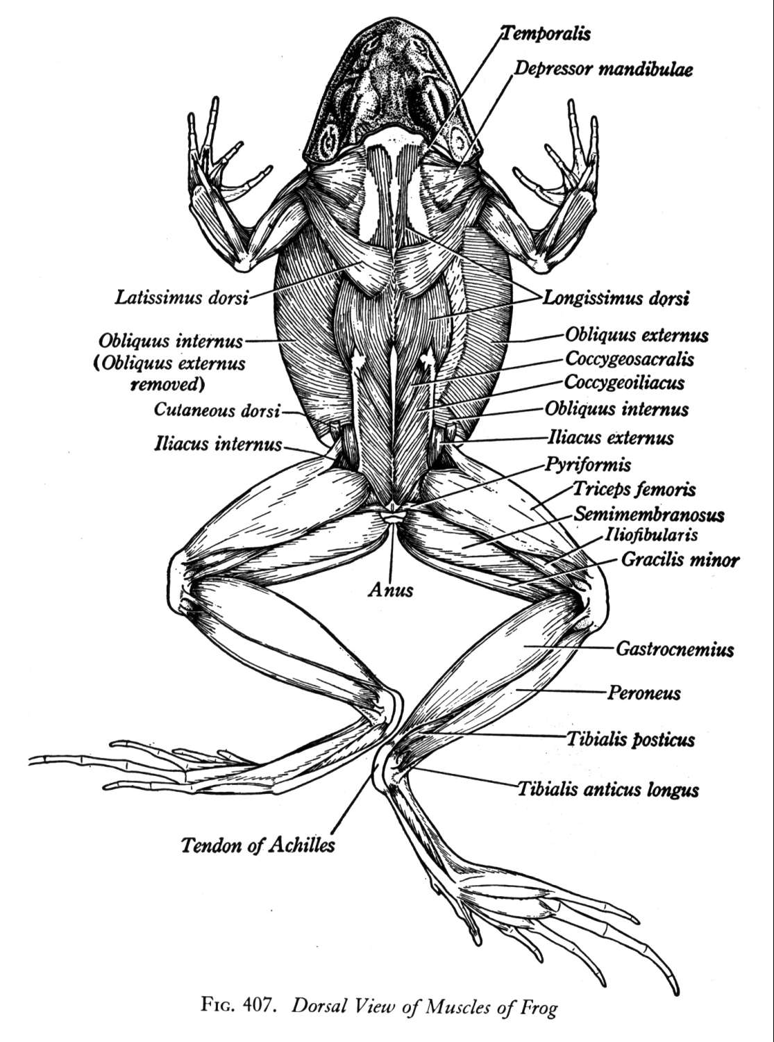
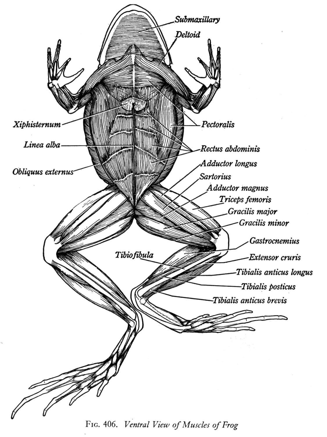
Skinning the Frog: Once you have identified the external features, you are now ready to remove the skin from the frog. To do this you must make small incisions that are just deep enough to cut through the skin. DO NOT CUT TOO DEEP OR YOU WILL DAMAGE THE UNDERLYING MUSCLES WHICH YOU NEED TO VIEW. Once you have removed the skin, use the diagrams below to help you identify the muscles of the frog.
After you have removed the skin, look at the inside of the skin you just removed. It is should contain many blood vessels. The frog depends upon its skin as a means for getting oxygen while submerged in the water. They are able to absorb the oxygen from the water through its skin. These blood vessels will carry that oxygen to the rest of the body. This type of "breathing" is known as cutaneous breathing.
Other Skin Characteristics:
Moist skin: Since they use the skin as an organ of respiration, the skin must be kept moist in order for gases to be exchanged. If the skin dries out, the frog would no longer be able to keep up with its oxygen demands because the lungs are not very efficient.
Smooth skin: Unlike fish and reptiles, the frog skin lacks scales. This gives the skin a smooth texture.
Mucus glands: Glands that secrete a thin, clear liquid that coats the skin and keeps it moist.
Granular glands: Glands that secrete a milky, alkaloid based substance. As with most frogs, the North American bullfrog secretions from the granular glands are not very toxic. These secretions just gives the them a bitter taste. The toxicity level of these toxins in the poison dart frogs, however, is off the charts. In some, the toxin from one tiny frog the size of your thumbnail might be enough to kill 10 grown men.
Turn the frog on its back and pin down the legs. Cut the hinges of the mouth and open it wide. Use the diagram below to locate and identify the structures inside the mouth. Use a probe to help find each part: the vomerine teeth, the maxillary teeth, the internal nares, the tongue, the openings to the Eustachian tubes, the esophagus, the pharynx, and the slit-like glottis.
Look for the opening to the frog’s cloaca, located between the hind legs. Use forceps to lift the skin and use scissors to cut along the center of the body from the cloaca to the lip. Turn back the skin, cut toward the side at each leg, and pin the skin flat. The diagram above shows how to make these cuts
Lift and cut through the muscles and breast bone to open up the body cavity. If your frog is a female, the abdominal cavity may be filled with dark-colored eggs. If so, remove the eggs on one side so you can see the organs underlying them.
Use the diagram below to locate and identify the organs of the digestive system: esophagus, stomach, small intestine, large intestine, cloaca, liver, gallbladder, and pancreas. Interestingly, the name of the large intestine does not refer to its length (the small intesting is several times longer than the large intestine), instead it refers to its diameter. The large intestine
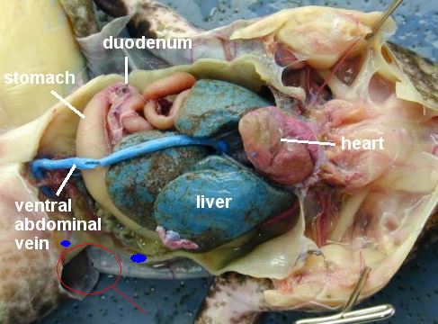
Again refer to the diagram below to identify the parts of the circulatory and respiratory systems that are in the chest cavity. Find the left atrium, right atrium, and ventricle of the heart. Find an artery attached to the heart and another artery near the backbone. Find a vein near one of the shoulders. Find the two lungs.
Use a probe and scissors to lift and remove the intestines and liver. Use the diagram on the next page to identify the parts of the urinary and reproductive systems. Remove the peritoneal membrane, which is connective tissue that lies on top of the red kidneys. Observe the yellow fat bodies that are attached to the kidneys. The fat bodies provide two important functions for the frog: 1) they provide a stored food source and 2) they provide protection for the delicate ovaries in the female frog. That is why both sexes will have fat bodies, but more times than not, you will see quite a bit more in the female frog.
Think Question: What time of year would you observe the largest amount of fat bodies in a frog in the wild? Answer: In the fall just before they begin to hibernate. This extra fat is what they will use to survive on for the extending winter while they are hibernating.
Find the ureters; the urinary bladder; the testes and sperm ducts in the male; and the ovaries, oviducts, and uteri in the female.
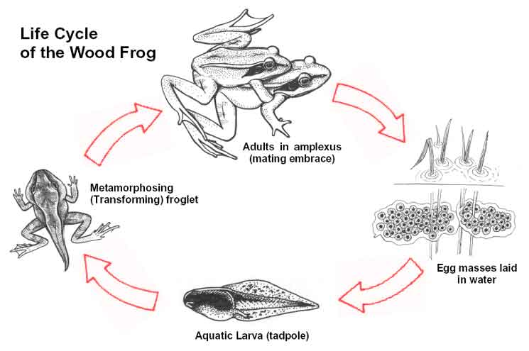
The frog life cycle is typical of all amphibians. After mating the eggs are laid in the water because they are NOT amniotic eggs. After hatching the tadpole emerges. They have gills like fish and are totally dependent on the water for all resources. As time goes on the tadpole will slowly start to develop the adult features. This "changing process" is known as metamorphosis. Once the frog develops its adult features it can leave the water for short periods of time, find a mate, and the process begins all over again. See the diagram below for the frog life cycle.
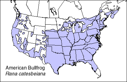
The North American bullfrog is native to much of the United States as seen in the diagram below. Unlike frogs that live in the desert, the North American bullfrog will hibernate to survive in the climate areas in the north. Because the weather is not too hot in the southeastern part of the US the frog does not hibernate, nor does it undergo estivation. It will be seen year round in these areas. You can see that it will not be found in the far north or mountainous areas. The necessary habitat of swampland is too scarce.
Dispose of your materials in the proper place..
Clean up your work area and wash your hands before leaving the lab. Complete your Frog Dissection Review on JupiterEd.
Click HERE to view a review of the frog dissection and information about what you need to know for the dissection quiz.