Shark Lab
Activity 3: Respiratory System
Click on any photograph for an enlarged view in a separate window.
Click HERE to access the Activity 3 Dissection Booklet
|
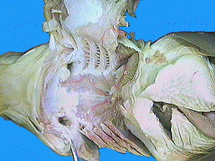
Examine the photographs of the gill pouches of the spiny dogfish shark by clicking the blue lettered links in the column to the right. The specimen in the photograph was prepared by cutting across the gill slits from the pectoral girdle to the corner of the mouth. It was cut across the ventral musculature to lay the entire preparation flat. The gills are the respiratory organs of the shark. They are composed of gill lamellae, blood vessels, and supporting cartilaginous structures are are located in a series of pharyngeal pouches. |
|
|
Examine the photographs of the inside of the oral and pharyngeal cavities of the spiny dogfish shark by clicking the blue lettered links in the column to the right. The oral cavity is the area enclosed by the jaws (mandibular arch) and the cartilage of the throat (hyoid arch). The triangular sharp teeth are arranged in several rows beginning at the outer edges of the upper and lower jaws. Behind the functional teeth are additional rows folded downward ready to replace any that are lost. The tongue of the shark is practically immovable and without muscles. It is supported anteriorly and posteriorly by cartilage. The pharynx is the portion of the alimentary canal posterior to the hyoid arch between the gills. Posteriorly it narrows to form the esophagus. The spiracles are openings in the anterior roof of the pharynx. The shark can bring water into its pharynx to the gills by way of the spiracles even when the shark's mouth is closed. |
|
|
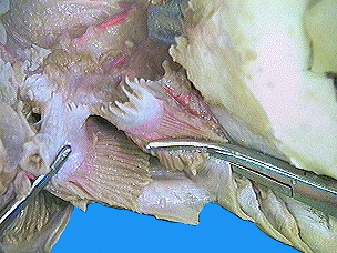
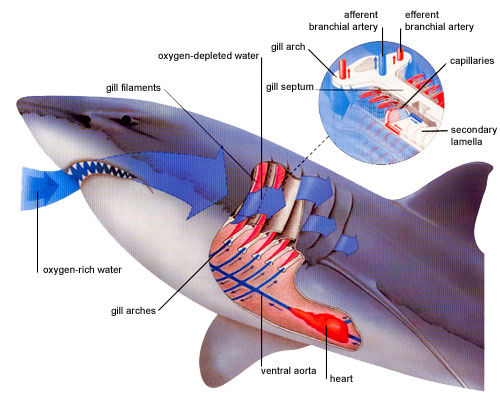
Examine the bottom view photographs of the shark's pharynx and heart by clicking the blue lettered links in the column to the right. The gill arches are the bony or cartilaginous curved bars on either side of the pharynx (throat) that support the gills of the shark. The gills are provided with a rich blood supply. Arteries run directly from the nearby heart to the gills bringing deoxygenated blood into the gill lamellae. It is here (at the gill lamellae) where the oxygen diffuses from the ventilating water current flowing over the gills into the blood. The gill lamellae have lots of capillaries to aid in the exchange |
|
|
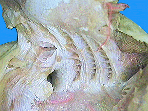
Examine the photographs of the shark's internal gill slits by clicking the blue lettered links in the column to the right. As you look at the pharynx you will see five internal gill slits. They lead into cavities called gill pouches, which lead to the outside by external gill slits. The gill slits are supported by cartilaginous gill arches and guarded by small cartilaginous papillae-like gill rakers which act as strainers to prevent food particles from leaving the pharynx through the gill slits. |
|
|
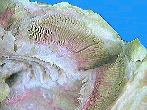
Examine the photographs of the shark's gill pouch by clicking the blue lettered links in the column to the right. The partitions between gill pouches are referred as branchial bars. The gill lamellae on one side of a branchial bar are called a demibranch, or half gill. The demibranchs on the anterior and posterior surface of a single branchial bar are termed a holobranch, or complete gill. Thus, one holobranch belongs to two different gill pouches; the anterior half (demibranch) to the anterior gill pouch, the posterior gill demibranch to the posterior gill pouch. |
|
|
Examine the photographs of the shark's gill lamellae by clicking the blue lettered links in the column to the right. The Gill Lamellae are radially folded, highly vascularized tissue attached to the surface of a tough connective tissue, the interbranchial septum. Each septum is attached medially to a portion of the cartilaginous gill arch. The superficial constrictor muscles act as flap-like valves to open and close the external gill slits. |
|
|
The diagram on the right shows how the shark's respiratory structures are able to remove the oxygen from the water. Notice how the incoming water is oxygen-rich, while the water exiting the gills is oxygen-depleted. |
|