Shark Lab
Activity 4: Circulatory System
Click on any photograph for an enlarged view in a separate window.
Click
HERE to access the Activity 4
Dissection Booklet
|
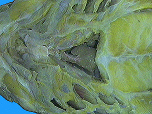
Examine the bottom view photographs of the skinned spiny dogfish shark by clicking the blue lettered links in the column to the right. The specimen in the photographs was prepared by removing the skin and the ventral musculature over the pericardial cavity. A membrane was removed to expose the heart and some of its major blood vessels. The pericardial cavity is the upper portion of the body cavity. It is much smaller than the lower cavity, which contains the digestive organs. The pericardial cavity is located anterior to the transverse septum and contains the heart and the major blood vessels leading to and from the heart. The pericardium is the membrane lining the inner walls of the pericardial cavity. |
|
|
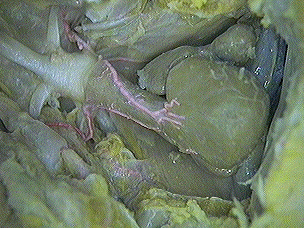
Examine the photographs of the shark's heart by clicking the blue lettered links in the column to the right. The ventricle is the thick muscular walled cavity that pumps blood through the conus arteriosus to the gills and the body. The conus arteriosis contains a series of semilunar valves that direct the blood flow. The atrium is thin-walled with two lateral bulging lobes. It pumps blood to the dorsal ventricle. |
|
|
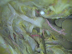
Examine the bottom view photographs of the shark's heart and cardinal sinuses by clicking the blue lettered links in the column to the right. Blood enters the heart through the sinus venosus which drains into the atrium. The posterior cardinal sinuses receive blood from the posterior parts of the body and drain through the common cardinal veins into the sinus venosus. |
|
|
Examine the photographs of the shark's ventral aorta by clicking the blue lettered links in the column to the right.The specimen in the photographs was prepared by removing the ventral hypobranchial muscles and connective tissues until reaching the lower jaw. The conus arteriosus was traced anteriorly following the major branching blood vessels. The anterior end of the conus arteriosus continues foward as the ventral aorta. It gives off five pairs of afferent branchial arteries which carry deoxygenated blood from the heart to the gills. The afferent branchial arteries pass laterally from the medial ventral aorta carrying deoxygenated blood to the gills. These afferent vessels enter the interbranchial bars and serve the holobranchs of the gill arches. |
|
|
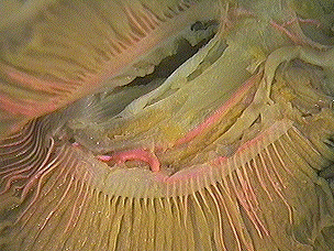
Examine the photographs of the male shark's efferent branchial arteries by clicking the blue lettered links in the column to the right. The specimen in the photographs was prepared by removing the mucous membrane from the roof of the mouth and pharynx. The efferent branchial arteries serve to return oxygenated blood from the gills. This blood is then distri bused to all parts of the body. Four pairs of arteries may be seen arising from the gills and uniting in the midline to form the median dorsal aorta. The efferent branchial arteries give off many branches. These carry oxygenated blood to the more anterior parts of the shark's body. The four pairs of efferent branchial arteries join at the dorsal midline to form the large dorsal aorta. The dorsal aorta passes posteriorly bringing oxygenated blood from the gills to virtually every part of the shark's body. |
|
|
Examine the photographs of the female shark's efferent branchial artery by clicking the blue lettered links in the column to the right. The specimen in the photographs was prepared by carefully dissecting to reveal the source of each efferent branchial artery in the gill lamallae of the gill pouches. The efferent collector loops encircle each of the first four gill pouches. |
|
|
Examine the photographs of the shark's collector loop by clicking the blue lettered links in the column to the right. Adjacent collector loops are connected to one another by branches which pass through the interbranchial septa. Below is a list of arteries and the organs or tissues they supply blood to:
|
|
| Portal
Circulatory Routes
A portal circulatory route is a venous system that begins as capillaries in one organ, follows veins to another organ and ends as capillaries in that other organ. In simple terms, it is a detour of the venous blood before it returns to the heart. The shark has two basic portal circulatory routes in which the blood is detoured from its normal route: 1. Hepatic Portal Circulation- this detour begins as capillaries in the digestive system (specifically near the intestinal area) and ends as capillaries in the liver. The blood needs to go to the liver for two specific reasons: 1) as the sharks food is digested, the nutrients need to go to the liver to be converted into the oils that the shark uses for energy. The glucose molecules will undergo metabolic changes in order to be converted into the oils, and 2) the liver acts as a detoxifying organ. Any blood toxins will be removed by the liver before the blood goes back to the heart. If this process didn't occur, the toxins would be redistributed again throughout the body.
2. Renal Portal Circulation- this detour begins as capillaries in the tail and end as capillaries in the kidneys. The purpose of the blood going to the kidneys is for filtration purposes. The waste products of normal cell activity need to be removed from the blood before it reaches the heart. The kidneys will act like large filters and remove impurities such as urea, uric acids, ammonias, etc. from the blood. These materials are toxic to cells and if redistributed along with the blood again to the cells the results could be fatal. Renal portal circulation ensures the removal of these toxic waste products BEFORE the blood gets redistributed.
|
Click here to see a diagram of the hepatic portal circulatory route
Click here to see a diagram of the renal portal circulatory route |