Sea Star Dissection Lab
If you missed the class period in which we dissected the sea star, you will need to view the following videos to help you understand what you missed in class.
Introduction:
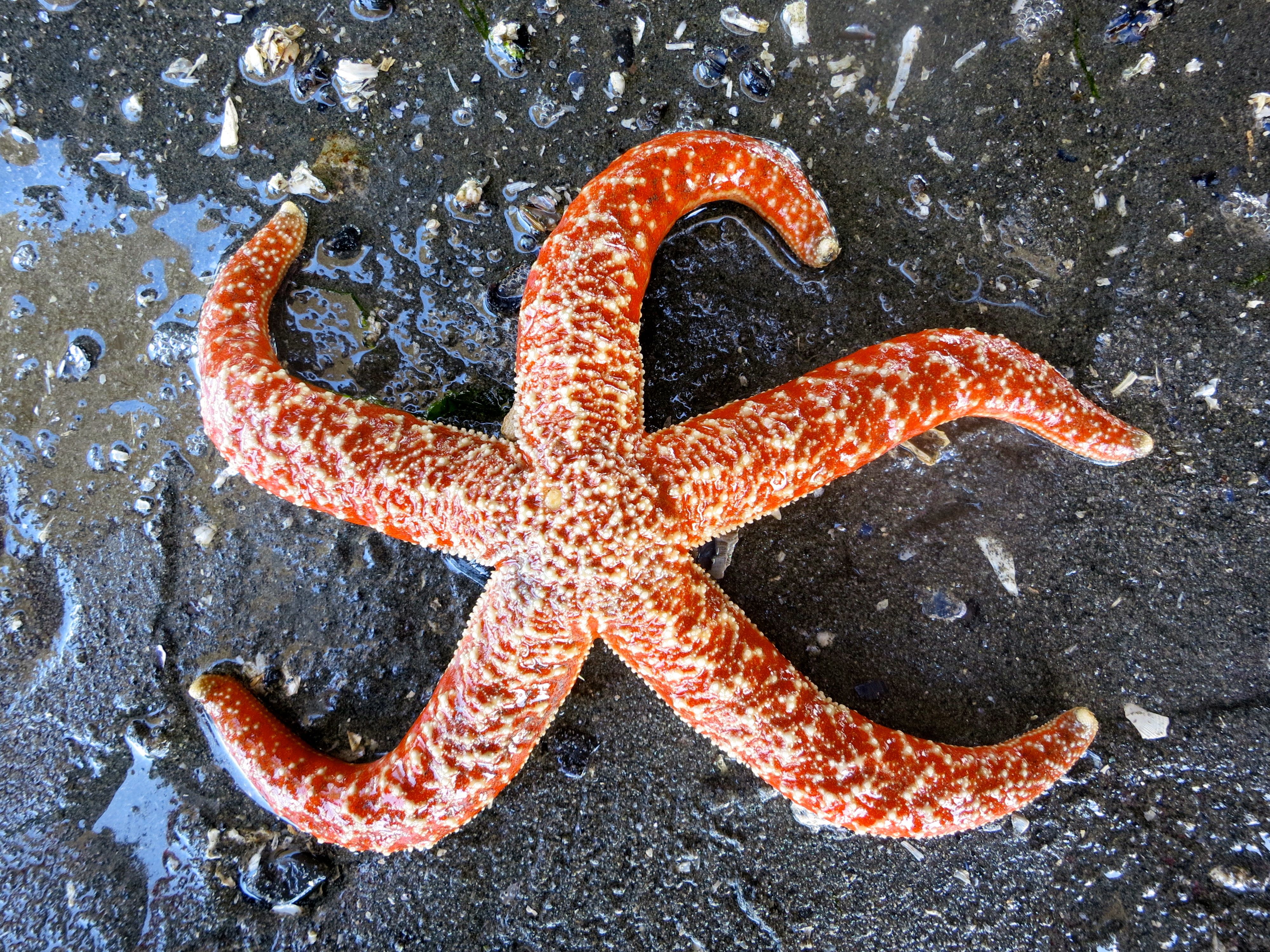
Echinoderms are radially symmetrical animals that are only found in the sea (there are none on land or in freshwater). The word "echinoderm" means "spiney-skin" in Greek. Many, but not all, echinoderms have spiney skin. There are over 6,000 species of echinoderms. Echinoderms usually have five appendages (arms or rays), but there are some exceptions.
Radial symmetry means that the body is a hub, like a bicycle wheel, and tentacles are spokes coming out of it (think of a starfish). As larvae, echinoderms are bilaterally symmetrical. As they mature, they become radially symmetrical. Most adult echinoderms live on the bottom of the ocean floor. Many echinoderms have suckers on the ends of their feet that are used to capture and hold prey, and to hold onto rocks in a swift current.
KINGDOM: Animalia
PHYLUM: Echinodermata
CLASS: Asteroidea
ORDER: Forcipulatida
FAMILY: Asteriidae
GENUS:
Asterias
SPECIES: forbesi
Therefore the scientific name for the common sea star is: Asterias forbesi.
Sea Stars:
Sea stars (group name Stelleroidea) are sometimes called starfish, though they are not real fish (they lack both vertebrae and fins). There are two sub-types of sea stars:
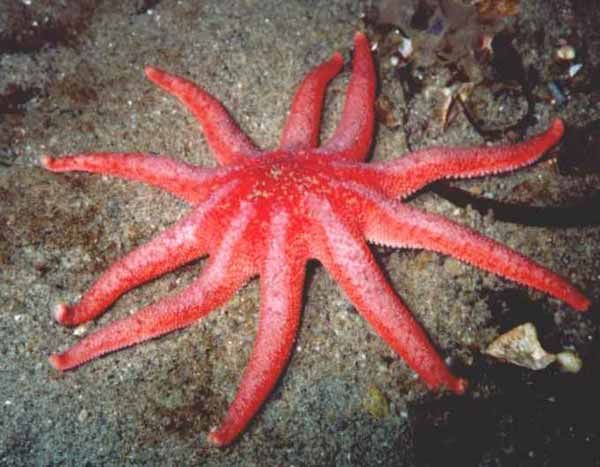
Asteroideas: these are the true sea stars and sun stars
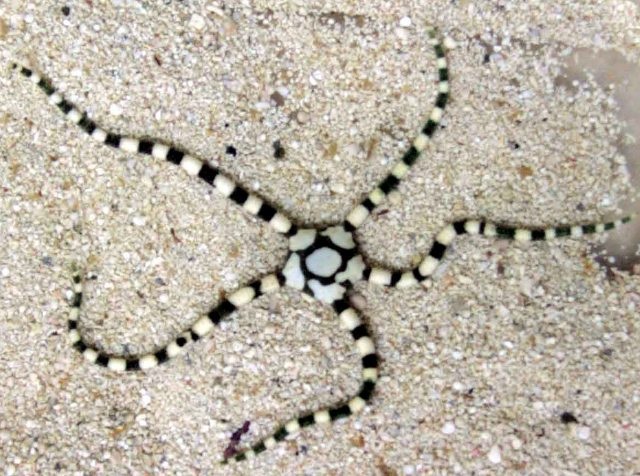
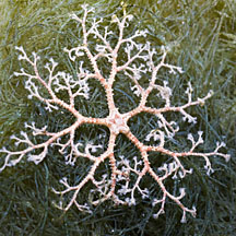
Ophiuroideas: these are the brittle stars and basket stars
| Asteroideas |
 |
| Ophiuroideas |
 |
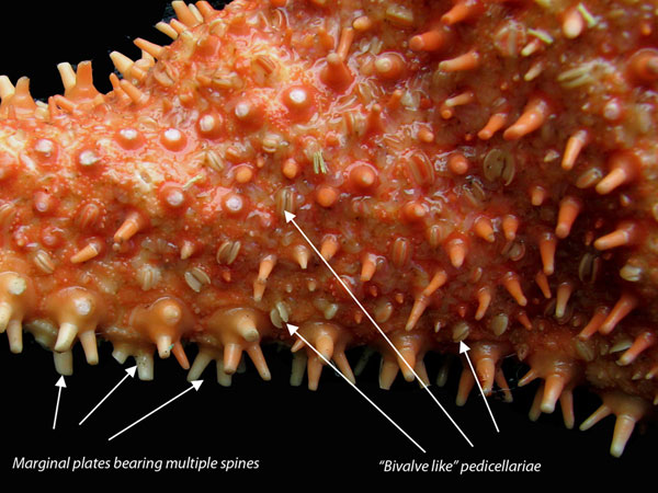
The differences between the two sub-types lies in how the arms connect to the central disk. Ophiuroids have rays that do not connect with each other. There is a distinct boundary between the rays and the central disk. Asteroids have rays that are connected to each other. Also it is harder to tell with asteroids where the central disk ends and the rays begin. The sea star's top surface (or skin) looks spiny if you examine it. If you look very closely you will notice that there are different types of growths on the surface. Some bumps are used to absorb oxygen, these bumps are called dermal branchiae. Pedicellaria are pincher-like organs used to clean the surface of the skin. Barnacle larvae could land on a sea star and start growing if it were not for these organs.
Pedicellaria
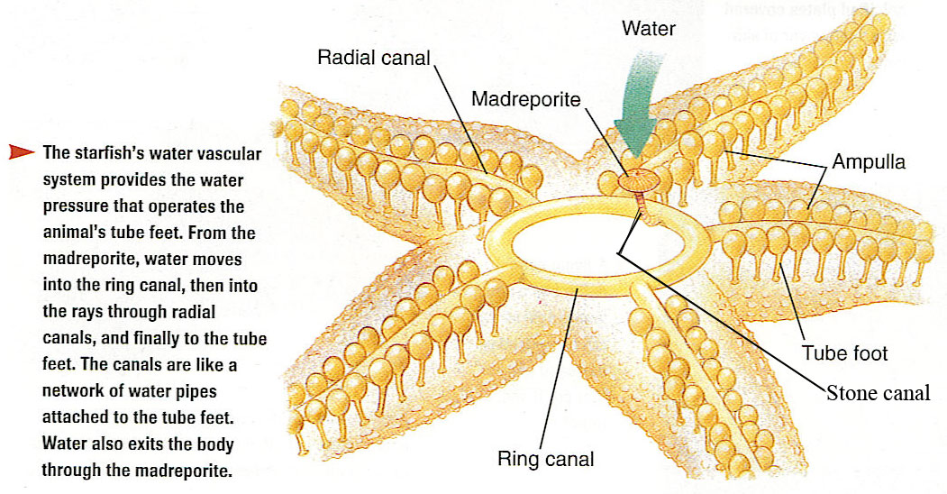
How do sea stars move?
Each sea star had
hundreds of tiny feet on the bottom of each ray. These are tube feet, or podia.
These tiny feet can be filled with sea water. The vascular system of the sea
star is also filled with sea water. By moving water from the vascular system
into the tiny feet, the sea star can make a foot move by expanding it. This is
how sea stars move around. Muscles within the feet are used to retract them.
Each ray of a sea star has a light sensitive organ called an eyespot. Though it
cannot see nearly as well as we do, sea stars can detect light and its general
direction. They have some idea of where they are going.
How do sea stars eat?
The diet of sea stars includes all types of mussels and other bivalves with clams and scallops being the most common meals for the sea star. The means of obtaining the food is quite unique in the animal kingdom. In order to make a meal out of a clam or scallop the sea star must use its tube feet as a way to "feel around" in order to find its food because they lack the ability to visually see. Once a clam has been designated for a meal, this is how the sea star will go about making a meal of a clam:
The sea star will use its rays and suckers on its tube feet to pry open the clam shell.
The sea star will then extend its stomach out of its mouth and into the clam's own shell.
The hepatic caeca will produce powerful digestive enzymes that will dissolve the soft body of the clam allowing the sea star to absorb this with its stomach.
When done feeding, the sea star will then use its retractor muscles to pull the stomach out of the clam and back into its mouth.
Sea Star Eggs & Scientific Research
Female sea stars produce large amounts of eggs and are readily available. Due to this, scientists have an almost endless supply of eggs available for research studies. Recently, scientists have studied the sea star eggs for their research in the areas of reproductive and development studies. The eggs (correctly called "oocytes") are harvested and stored in their pre-meiosis phase and then stimulated later to complete their division. This allows the scientists to study the development of the eggs at any point in the cell division. Scientists hope to learn more about the process of the cell division involved with the regenerative ability of the the sea stars.
Click HERE for a more detailed and comprehensive Dissection Manual. There will be numerous questions on the lab review that are directly from this source.
DISSECTION PROCEDURE:
1.
Place the starfish on your
dissection tray with its aboral surface facing upward.
2.
Using your diagram sheets and the
pictures below to locate the following structures, and note their functions:
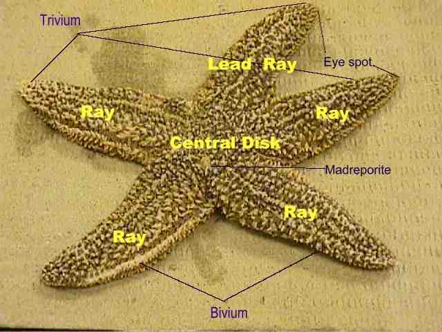
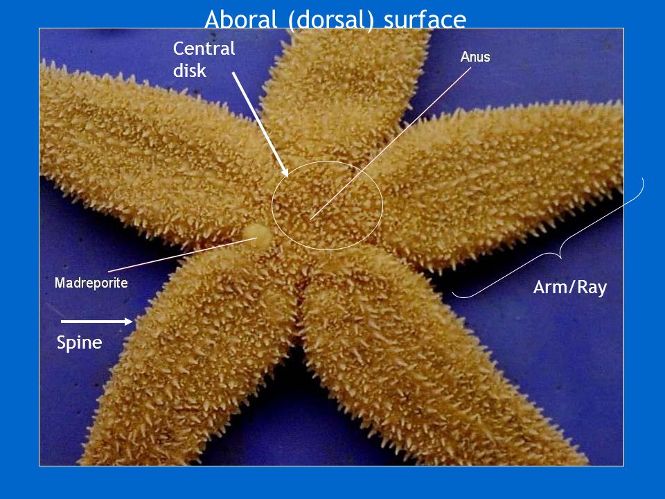
¨ Arms or Rays: these are the five extensions that you see projecting from the middle of the starfish. These are highly regenerative and are replaced easily when damaged. You might even see one of our starfish that has a ray that is significantly smaller than the rest of them. This is because the ray had been damaged or lost and it has regenerated a new one. The two rays which are closest to the madreporite are known as Bivium. The other three rays are referred to as the Trivium.
¨
Central Disk:
this is the middle area of the starfish from which the rays extend.
It is often poorly defined and difficult to locate
the perimeters, but on some you may be able to distinguish a pentagon shaped
area.
¨
Aboral Surface:
the aboral surface is the surface that does not contain the mouth.
¨
Madreporite: this
is a small, white, circular area that is located in the central disk area.
It is usually off-center and is sometimes called the
sieve plate.
It is used by the starfish to take in water to fill
its water-vascular system.
If you scratch it with your probe you will notice
that it is rather hard and feels like stone.
¨
Anus: the anus is
rather minute and difficult (pretty close to impossible!) to see but it is also
located on the central disk.
Wastes are excreted through this opening to the
outside of the starfish.
¨
Spines:
the entire aboral surface is covered with many short, rough, limy spines
¨
Eyespot:
the eyespot is located at the distal end of each ray.
It is a collection of photosensitive cells which the
starfish uses to detect light or absence of light.
3.
Now flip your starfish over so you
can view the oral surface.
Use your diagram sheets and the pictures below to
identify the following structures:
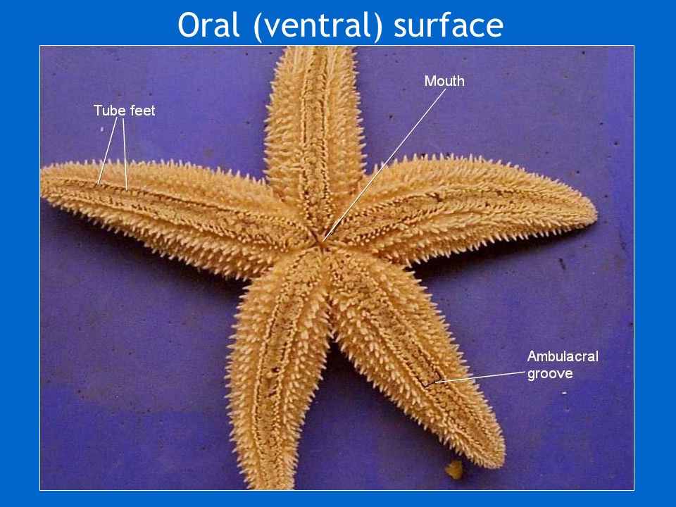
¨
Ambulacral Groove:
this is where the tube feet are located.
They are found along each ray.
¨
Ambulacral
Spines: these are slender rods located on the margins of the
ambulacral grooves.
¨
Tube Feet:
soft, slender, with expanded tips.
There are two or more rows in each ambulacral
groove.
¨
Mouth: opening in
the middle directly beneath the central disk where all the arms connect.
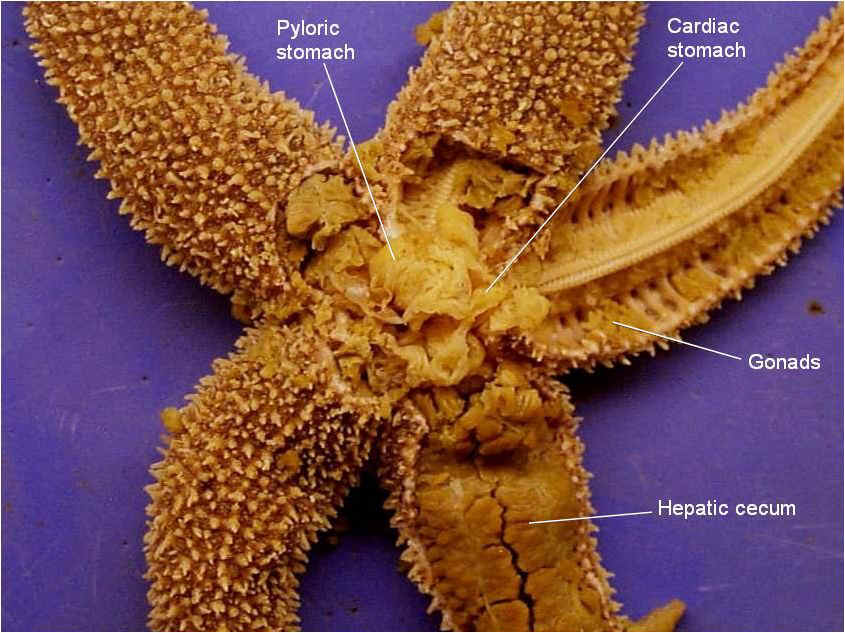
1. Use the scissors to cut off the extreme tip of each arm of the bivium. Then cut along the lines labeled A, B, and C as shown in the diagram below. Do this to both of the rays of the bivium.

2.
In turn, lift and carefully remove
the aboral surface of each arm, loosening the delicate mesentaries beneath by
which the soft organs are attached.
Also, cut around the central disk to expose the
stomach underneath.
3.
Use your diagram sheets and the
pictures below to identify the following structures:
¨
Coelom: space
containing the internal organs; lined with thin ciliated peritoneum
¨
Stomach:
sack-like structure found underneath the central disk.
¨
Retractor Muscles:
small, sinewy structures that connect to the stomach.
These are used to pull the stomach back into its
mouth once the starfish is done feeding.
¨ Hepatic caecum: long, greenish organs found in each ray. Has many finger-like lobes. These organs are used for secreting digestive juices and enzymes needed for feeding. The plural form of the word is "hepatic caeca".
¨
Gonads: small,
bi-lobed structures found below the hepatic caeca in each arm.
May be very small in some specimens due to the fact
that the starfish may not be sexually mature yet.
4.
In order to determine the sex of
your starfish, you must examine a small portion of the gonad with the
microscope.
5.
Make a mounted slide by taking a
SMALL PORTION of the gonad and placing it on the microscope slide.
Cover this with a cover slip and observe under the
low power objective.
6. If your starfish is a female you will see the eggs. This resembles small circular-looking objects. Below is a picture of what starfish eggs will look like under magnification.
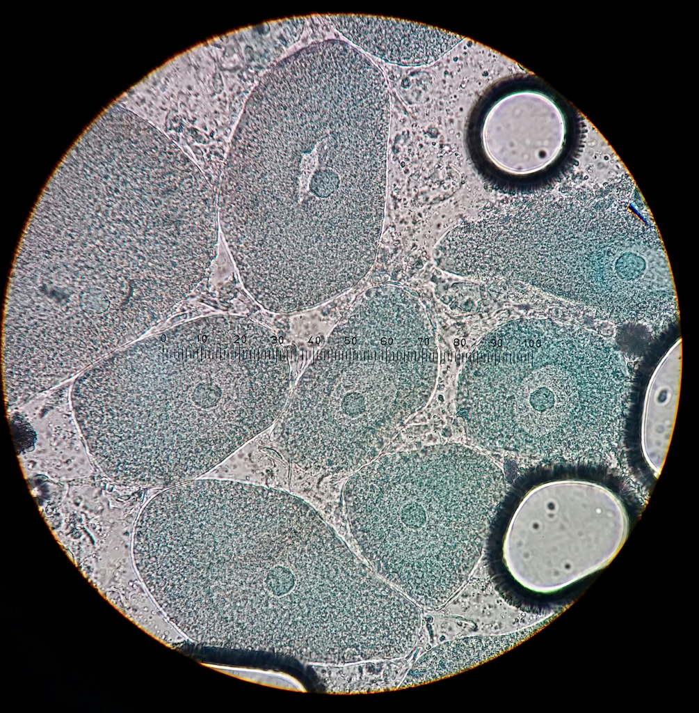
7. If your starfish is a male you will see the sperm. Instead of seeing circular objects you will see something that resembles sand. These are the sperm cells. Your specimen may not be as clear as the one below, in fact it may just look like tiny grains of sand......but starfish sperm look like the picture below.
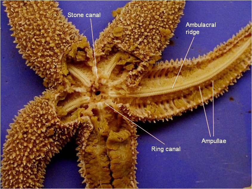
Part 3: Water Vascular System
1.
Remove the stomach from the central
disk area.
2.
You will now be able to see the
calcite skeletal system of the starfish and also the parts of its water vascular
system.
Use you diagram pages to locate the following structures:
¨
¨ Tiedemann bodies: nine, small swellings in the ring canal.
¨
Ampullae: many,
small, spherical structures in the floor of the coelom.
These connect to the tube feet.
¨
Tube feet
3.
Dispose of your starfish in the
garbage and clean up your trays and utensils.
4. Complete your lab review worksheet. Click HERE for the Starfish Dissection Lab Companion that will detail what you will need to know for the quiz.