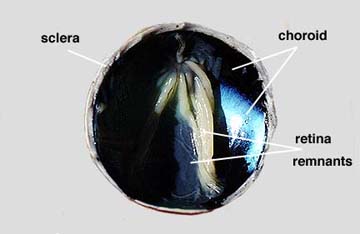

Step 6: Use your forceps to peel the retina away from the underlying choroid coat. The retina should remain attached at the blind spot. The choroid coat is dark and relatively thin. Use your forceps or probe to gently separate the chorid from the outer sclera. Verify that the eye has three distinct layers, the retina, choroid and sclera. See left photograph above. The choroid contains an extensive network of blood vessels that bring nourishment and oxygen to itself and the other two layers. The dark color, caused by pigments, absorbs light so that it is not reflected around inside of the eye. The tapetum lucidum, which functions to reflect light onto the retina. It especially helps animals with night vision since it can reflect light even at very low intensities. The tapetum lucidum in the sheep eye is shiny, glittering with a bluish color. Humans and other primates do not have this structure. Many of the animals that do have the tapetum lucidum are nocturnal animals or animals that occupy the deep ocean depths. They use this for vision in extremely low light regions.
In just a moment you will see that the choroid extends forward to the ciliary body. Take the notes you need to record what you have observed so far.
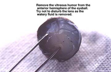
Step 7: Use your forceps and probe to remove the vitreous humor from the anterior hemisphere of the eye. See right photograph above. This will take some time and effort as the semi-fluid material separates easily. It helps to turn the hemisphere on edge and to use a scrapping motion to remove the fluid. Try not to disturb the lens that is just below the vitreous humor. Take the notes you need to record what you have observed so far.