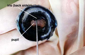
Step 10: When the lens is removed, an opening, allowing light to enter the eye is seen. This opening, the pupil is located in the center of the iris. Two muscle layers of the iris regulate the size of the pupil. One layer increases the pupil size with decreasing light intensity and the other layer reduces pupil size with increasing light intensity. Note the oblong shape of the sheep pupil, in humans the pupil is circular. The back side of the iris can be seen just above the pointer in the photograph. Part of the iris is being lifted by the pointer but the iris continues all the way around the pupil opening.
A second cavity or space is present between the iris and the cornea. This space is filled with a second semi-liquid fluid, the aqueous humor. This fluid, like the vitreous humor helps to maintain the shape of the eye. Glaucoma is a condition where the fluid pressure becomes too high causing eye damage. Take the notes you need to record what you have observed so far.