Chicken Wing Dissection
|
|
Human Anatomy Chicken Wing Dissection |
|
Background Information
Chicken wing dissections are conducted to
explore the structure and function of muscles, bones and joints, which are
comparable to that of a human arm, they have many of the same structures due to
their shared evolutionary history as vertebrates. Skeletal muscles are
attached to bones, give shape to the body, generate heat, and make movement
possible. Skeletal muscles cannot function without the bones of the
skeletal system. Muscles pull on the bones in specific ways and with the
guidance of ligaments allow joints to flex or extend in a specific direction.
The skeletal system is a network of various living tissues, which provide
protection for organs and give the human body its structure. It is also
the site of blood formation. In this activity you will study
chicken wing structure and function, which is comparable to that of the human
arm.
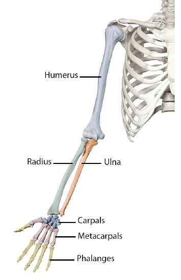
The Human Upper Limb Anatomy
The arm, as defined,
reaches from the shoulder to the wrist.
It consists of two basic parts:
1.
The upper arm:
which extends between the shoulder and the elbow
2.
The lower arm:
which extends between the elbow and the wrist
The upper arm is formed by one long bone, the
humerus.
The proximal end of the humerus is rounded and fits into the cup-shaped
depression on the scapula known as
the glenoid cavity. Together they
form the ball-and-socket joint of the shoulder which allows for a circular
movement known as circumduction.
The two bones of the
forearm are the radius and the
ulna.
The ulna is in a fixed position, but the radius can rotate over the ulna.
This makes rotation of the forearm possible in motions such as twisting a
screwdriver.
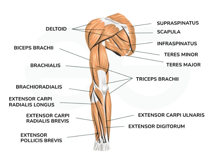
Skeletal muscles
are responsible for hundreds of movements.
When an organism wants to move, signals travel from the brain to the
skeletal muscle cells. The muscle
cells then contract, or get shorter.
Strands of tough
connective tissue connect the
skeletal muscles to the bones.
These strands of tissue are called
tendons. When a muscle that
connects two bones gets shorter, the bones are pulled closer to each other.
For example, tendons attach the biceps muscle to a bone in your shoulder
and to a bone in your forearm. When
the biceps muscle shortens (contracts), your forearm bends toward your shoulder.
The skeletal muscles often
work in pairs to produce smooth,
controlled motions by pulling, or contracting.
When one muscle in the pair bends part of the body, the other muscle
extends or straightens part of the
body.
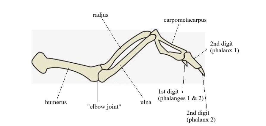
Bones of the
Chicken Wing
The upper wing consists of the
humerus, which is at one end, and the
ulna and the
radius at the other end which forms
the lower wing.
These bones connect at the elbow
joint. The rest of the wing is
composed of the carpals and
phalanges, which are modified, but
still present, just like in the human wrist and hand.
Glossary of Terms Associated with the Skeletal & Muscular Systems
biceps muscle: a two-headed muscle which is one of the chief flexors of the forearm that lies on the upper arm between the shoulder and the elbow
bone marrow: the soft blood-forming tissue that fills the cavities of large bones containing fat and blood cells
bones: the main material that forms a vertebrate skeleton, principally collagen fiber and calcium phosphate
capillaries: small blood vessels located within the body tissues that connect small arteries to small veins; location where gas exchange takes place
cartilage: thick, slippery tissue that coats the ends of long bones where they meet to form a joint
connective tissue: supports and binds other tissues of the body
humerus: the long bone of the human upper arm or of a forelimb in other vertebrate animals
joints: part of body where bones are connected
ligaments: bands of fibrous tissue that holds bones together in a joint
metacarpals: bones of the hand can be grouped into three categories: Carpal Bones, Metacarpals and Phalanges. The metacarpals and phalanges of birds are very heavily modified
muscles: tissue that can undergo repeated contraction and relaxation, so that it is able to produce movement of body parts
phalanges: bones of the fingers and toes, phalanges of birds are very heavily modified
radius: one of the two large bones of the forearm, the other being the ulna
tendons: elastic tissue that attaches muscles to the bones
triceps muscle: large muscle on the back of the upper limb of many vertebrates responsible for extension of the elbow joint (straightening of the arm)
ulna: the long bone found in the forearm that stretches from the elbow to the smallest finger or phalanges
vertebrates: an animal that has a backbone or spinal column
dissecting tray
raw chicken wing
dissection kit
latex gloves
Click the here for a video that details the chicken wing dissection.
![]()
Dissection Procedure:
1. Place the chicken wing on the dissecting tray. Study the external appearance and structure of the wing. Feel the skin that is covering the bones and look for places where the feathers were attached.
2. Identify the upper wing, the lower wing, and the wing tip. See Figure 1.
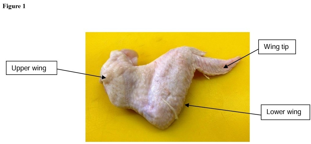
3. Feel for bones through the flesh, the upper wing consists of one long bone called the humerus; the lower wing consists of two bones, the radius and the ulna. The wing tip consists of modified hand bones, the metacarpals and phalanges. The most notable difference between the human anatomy and the bird anatomy is the fusion of some of the carpals and metacarpals into one bone called the carpometacarpus bone. Also the phalanges in the bird are fused to allow for the attachment of feathers.
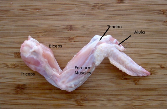
4. Feel for the muscles and tendons, there are two big muscles on the front and on the back of the upper wing that bend and straighten. These muscle are known as the biceps muscle and triceps muscle. Tendons attach these muscles to the bone in the shoulder and the bones in the forearm and lower wing.
5. Examine the wing at the shoulder joint where it was removed from the body. You should be able to see the slipper shiny white cartilage covering the end of the bone. This is known as hyaline cartilage. Its function is to cover and protect the bones and allow for smooth movements within the joint capsule. Further examination should reveal the shiny white ligaments that connected the bone to the joint.
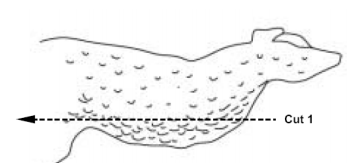
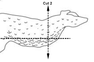
6. Carefully cut the skin of the wing down its length using the dissection scissors (Cut 1). Make a T-shaped cut (Cut 2) as seen in the diagrams below.
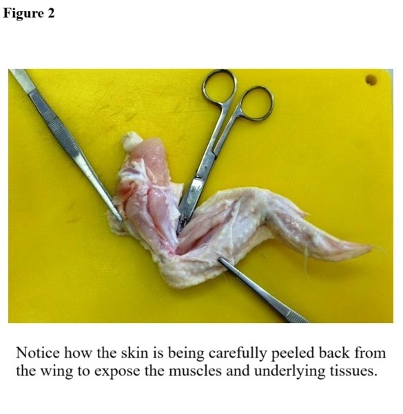
7. Use the foreceps to gently peel back the skin without damaging the underlying tissues, starting with the cut end and working down towards the wing tip. Cut and gently scrape the skin free from the muscle underneath using the dissection scissors. See Figure 2.
8. Look at the fatty tissue on the underside of the skin. The fat is yellow in color and should feel greasy. Notice the capillaries and muscles that are surrounded by connective tissue, which appears as a thin film or membrane.
9. Examine the exposed skeletal muscles of the wing. They appear as pink bundles of fibers. These muscles are attached to the bones and cause movement of the bones when they contract and relax.
10. Flex the wing and observe what happens when you pull on the triceps muscle and the biceps muscle. Observe how the muscles work in opposing pairs to move the bones.
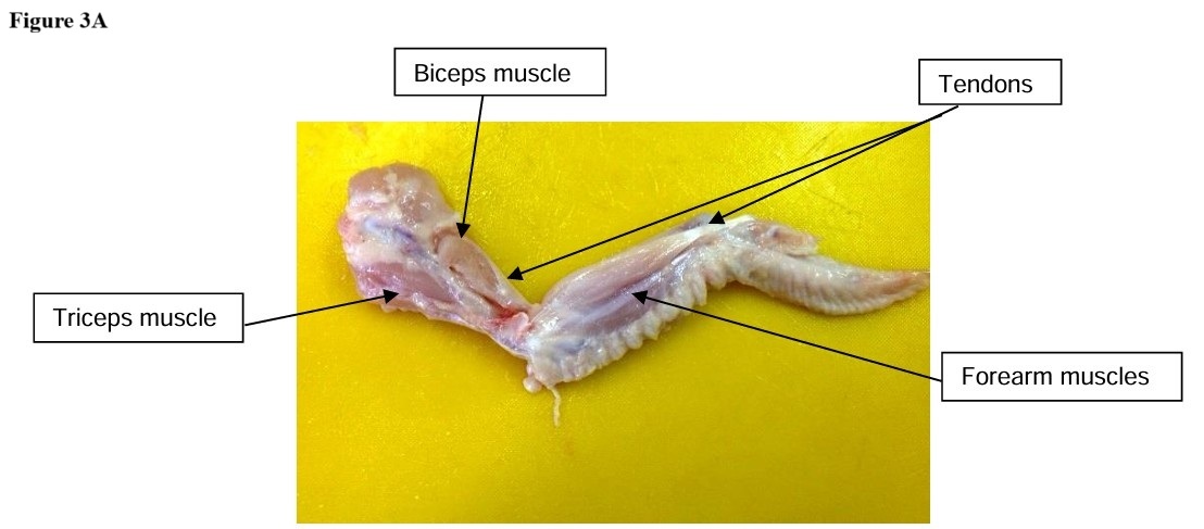
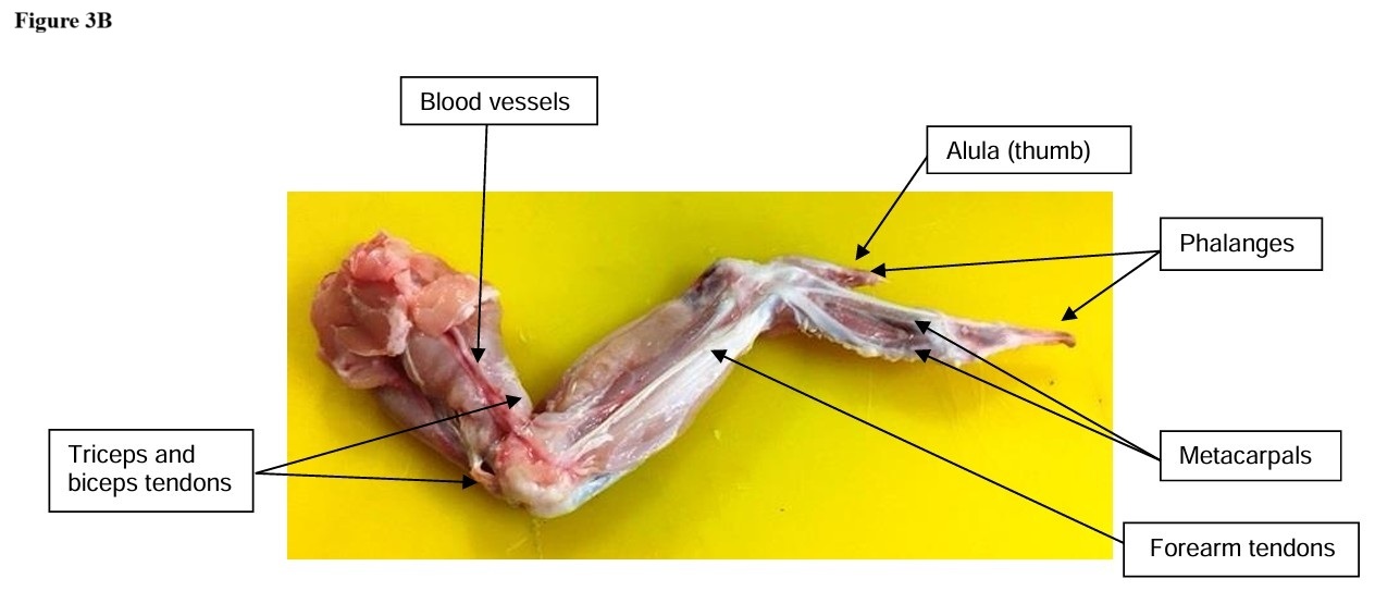
11. Look for the shiny white strands of tissue that attaches the muscles to the bones. These tissues are called tendons. See Figure 3A and 3B.
12. Move the wing again and explore how the muscles, tendons, ligaments, and cartilage play roles in the movement of the wing.
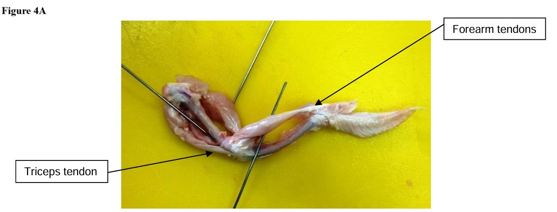
13. Cut the muscle and fat off the wing to expose the bone. Observe how the different bones of the wing work together. See Figures 4A and 4B.
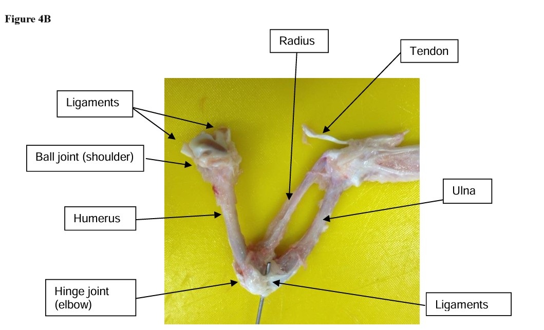
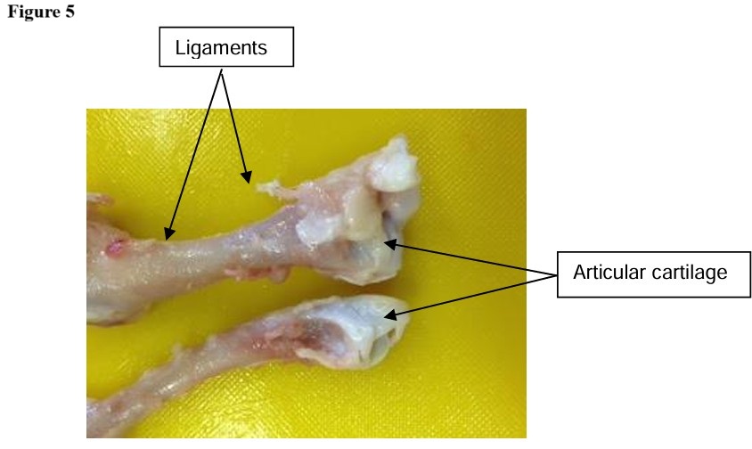
14. Observe the cartilage that covers the bones where they meet forming the joints, and locate the ligaments bonding the joints together. See Figure 5.
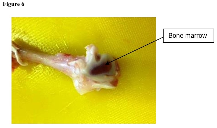
15. Remove the cartilage from the surface of the bone OR try to break one of the bones in the middle (use your hands for this.....DO NOT use a scissors or scalpal). Inside the bone you will see it is hollow and filled with a pinkish-red jelly-like material known as marrow. See Figure 6.
Clean-up Procedure:
1. Return all instruments that you used to the tray on the front lab table. These will be washed in the dishwasher to clean and sterilize them so that they will be safe to use for the next lab.
2. Place all chicken parts in the garbage receptacle as indicated by Mr. Breitkreutz. This will be disposed of in the garbage bins following class so that no cross contamination can occur in the classroom.
3. Wash your trays with soap and hot water to clean them. The final step of cleaning these items is to dip them in the bleach solution sink along the west wall of the classroom. This will disinfect the materials and make them safe for the next use.
4. Make sure to dry the mats and trays before returning them to the shelves.
5. Wash your hands with warm water and soap before leaving class.
Assignment:
1. Complete the Jupiter Ed assignment (Chicken Wing Dissection Lab Review) by the due date posted on the Human Anatomy website.