|
|
Sheep Eye Dissection Lab |
|
Purpose:
The anatomy of the human eye can be better shown and understood by the actual dissection of an eye. One eye of choice for dissection, that closely resembles the human eye, is that of the sheep. Sheep eyes are removed at the time the animal is slaughtered and then preserved for later use. Differences between the two eye types will be mentioned later in the lab as the dissection progresses.
Materials Needed:
preserved sheep eye
dissecting tray
dissecting kit
paper towels
Dissection Procedure:
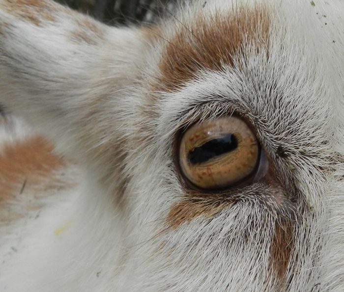
1. Wash the sheep eye under running water to remove the preserving fluids. Dry the eye with paper towels. Examine the front of the eye and locate the following structures on the eye: (use the picture below for assistance)
eye lid
cornea
sclera (white of the eye)
fatty tissues (adipose)
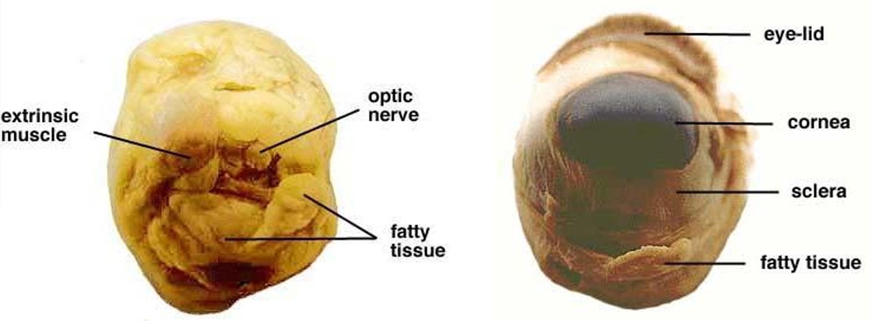
2. Examine the back of the eye and locate the following structures:
extrinsic muscle bundles
adipose tissues
optic nerve
3. The four extrinsic muscles (humans have six) move the sheep eye while the fatty tissues cushions the eye within the eye socket. If the optic nerve is not visible use the probe to move the fatty tissue around until the nerve is exposed.
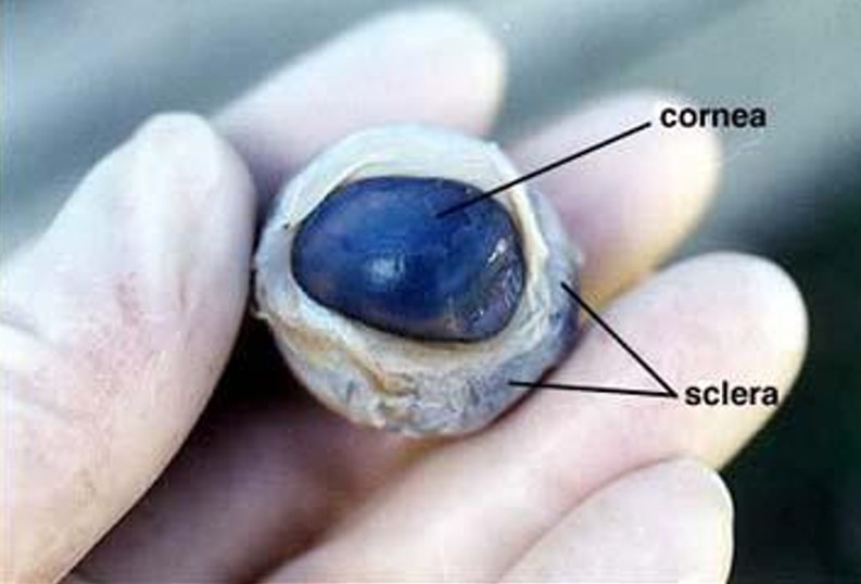
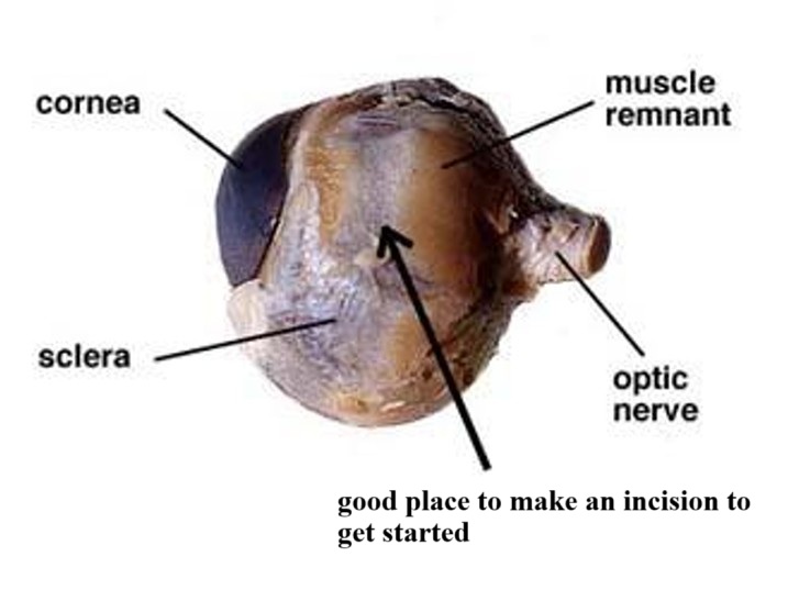
4. Use your scissors to cut away the eye lid, muscle and adipose tissues from both the front and rear surfaces of the eye. Be careful not to remove the optic nerve. Cut along the surface of the sclera until all the excess tissue is removed and your eye specimen looks similar to the pictures below:
 |
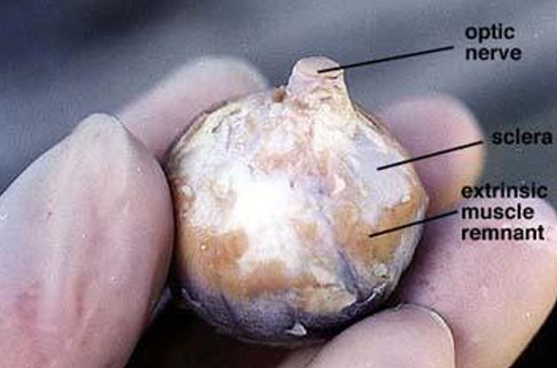 |
5. The sclera is very tough so you do not need to worry about cutting into this layer of the eye. When you have finished removing the tissue surrounding the eye, identify once again the following structures on your eye:
sclera
cornea
optic nerve
remaining remnants of the extrinsic muscles
6. One significant difference you may see on a preserved eye, like the one you are observing, and a living specimen is the color of the cornea. In a living specimen the cornea is clear and transparent to allow the light rays to enter the eye unimpeded. The cornea of the preserved eye is quite cloudy. This is due to the death of the tissues of the cornea.
7. Place some paper towels along the bottom of your dissection tray (this will help absorb some of the materials from inside the eye and make your cleanup a bit easier). Place your eye specimen in the dissection tray on top of the paper towels. Turn the specimen so that the cornea is on your left and the optic nerve is on your right. Select a place to make an incision of the sclera about midway between the corner and optic nerve. See the diagram below for assistance. Use the point of the scalpel to make a small cut through the sclera. Make sure to keep your mouth closed during this process. This statement would not be included in the lab unless past experiences made it necessary. Fluid should ooze out of the eyeball when you have cut deep enough. You will be reminded how tough the sclera is when you make this cut.
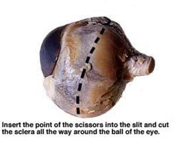
8. Insert the pointy end of your scissors into the slit made by the scalpel blade and cut the sclera with a shallow snipping motion. Turn the eye as you continue the cutting action. Cut the sclera all the way around the ball of the eye. You will need to support the eye in the palm of your hand while you complete this step of the dissection. The eye will be very slippery and cutting into it without holding securely in your palm will only make for a dangerous situation and you won't be able to cut it open properly. See the diagram below for assistance. The liquid that is coming out of the eye at this point is the aqueous humor. It is much less viscous than the vitreous humor.
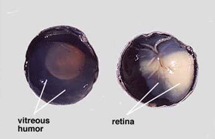
9. Arrange the two cut hemispheres of the eye as you see in the left photo below. Observe the gelatinous fluid. This is the vitreous humor that fills the central cavity of the eye. It is transparent in the living eye, but like the cornea, may be cloudy in the preserved specimen. The vitreous humor, along with the aqueous humor helps maintain the shape of the eye. The retina lines the posterior cavity of the eye and extends forward to the ciliary body. Use your probe to lift and pull the retina back from the underlying choroid layer. See the photo on the right side for assistance. Notice that the retina is only firmly attached to the choroid at one place. This region is the optic disc or "blind spot". Here the nerve fibers leave the retina and form the optic nerve which is directly behind the optic disc (blind spot).
 |
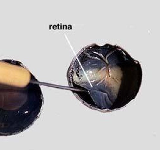 |
10. Use your tweezers to peel the retina away from the underlying choroid coat. The retina should remain attached at the blind spot. The choroid coat is dark and relatively thin. Use your probe to gently separate the choroid from the outer sclera. Verify that the eye has three distinct layers: the inner layer (retina), the middle layer (choroid), and the outer layer (sclera). See the picture below:
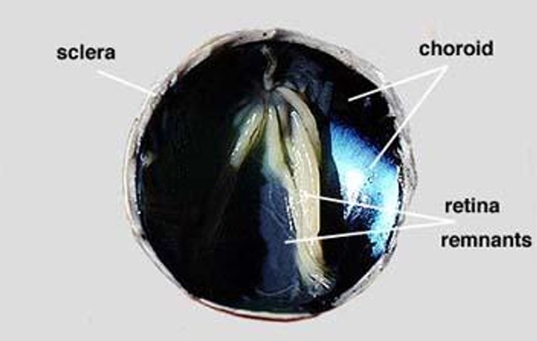
11. The choroid contains an extensive network of blood vessels that bring nourishment and oxygen to the choroid as well as the other two layers of the eye (sclera, retina). The dark color, caused by pigments, absorbs light so that the light is not reflected around the inside of the eye. This would result in a scattered light path and blurred vision.
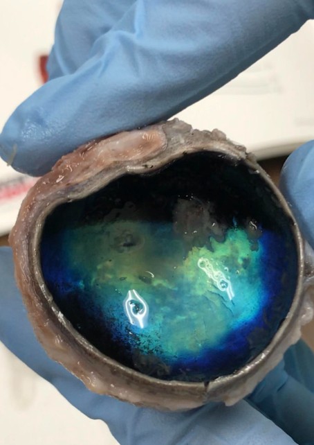
12. The tapetum lucidum, which functions to reflect light onto the retina, helps some animals see better at night. The reflective nature of this tissue layer can reflect light even at very low intensities. The tapetum lucidum in the sheep eye is shiny, and glitters with a greenish-blue color. See the picture below:
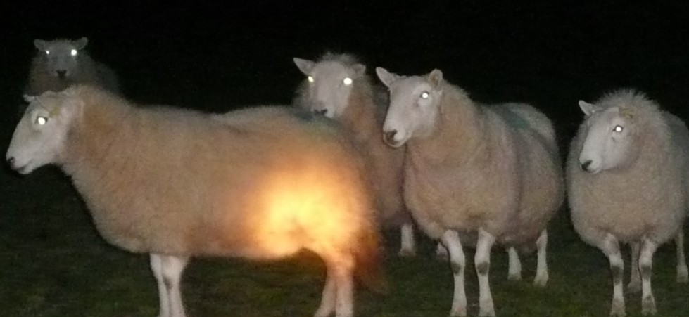
13. Animals that are nocturnal (animals that are active at night) often have the tapetum lucidum which helps them see better at night. Humans are diurnal (animals active during the day) and therefore do not need this structure. Sheep and other grazing animals are also diurnal, but have evolved these structures because they are MOST active at dawn and at dusk when light intensities are quite low which is quite an advantage for them. You know an animal has this structure if their eyes "shine" or "glow" at night when an artificial light is directed into their eyes as shown below:
 |
 |
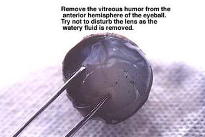
14. Use your tweezers and probe to remove the vitreous humor from the anterior hemisphere of the eye. See right photo below. This may take some time and effort as the gelatinous fluid separates easily. It helps to turn the anterior hemisphere on its side and scrape the remaining vitreous humor out. Often the lens will come out with the vitreous humor.
 |
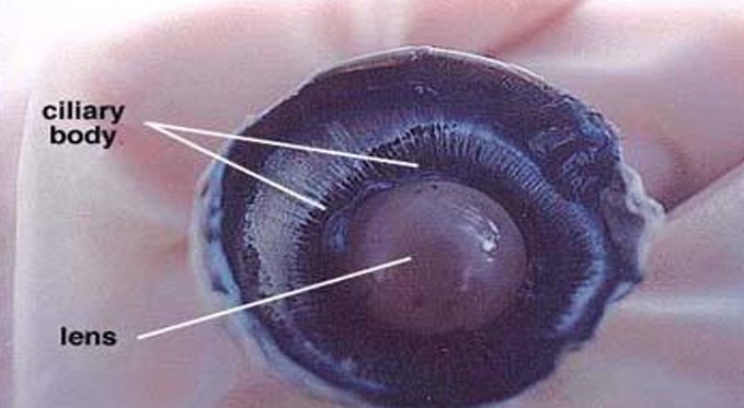 |
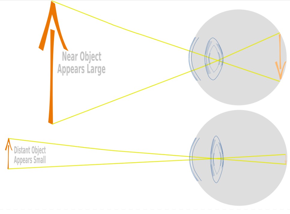
15. Removal of the vitreous humor reveals the lens, ciliary body of the iris, and suspensory ligaments. In the living animal, the lens is normally transparent, except when as a condition of aging, the lens turns cloudy. This cloudy condition is known as cataract and it can prevent or reduce the amount of light reaching the retina. Cataracts can be treated by removing the lens and replacing it with a stiff artificial one. The normal lens is convex shaped and somewhat elastic. It is held in place by suspensory ligaments that in turn join with the smooth muscle containing the ciliary body. When smooth muscle fibers contract the resulting force flattens the lens and the degree of refraction is reduced. In this case, the ciliary body is responsible for moving and shaping the lens for focusing. Relaxation of the smooth muscle results in a thickening of the lens and greater refraction of the light rays. See the diagram below for better understanding of how the lens works to see both near and far objects.
16. Remove the lens by pulling it free from its attachments (if it hasn't already detached). Note the shape of the lens, its stiffness and opaqueness. Keep in mind that in a living animal the lens is transparent and quite flexible. There may also be some small suspensory ligaments surrounding the edge of the lens.
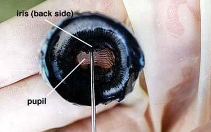
17. When the lens is removed, an opening, allowing light to enter the eye is seen. This opening is called the pupil and is found in the center of the iris. Two muscle layers of the iris regulate the size of the pupil. One layer increases the pupil size with decreasing light intensity, and the other layer reduces the size of the pupil with increasing light intensity. See the picture below:
 |
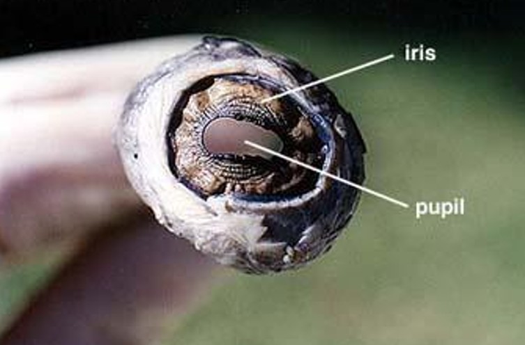 |
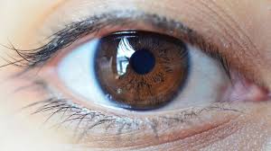
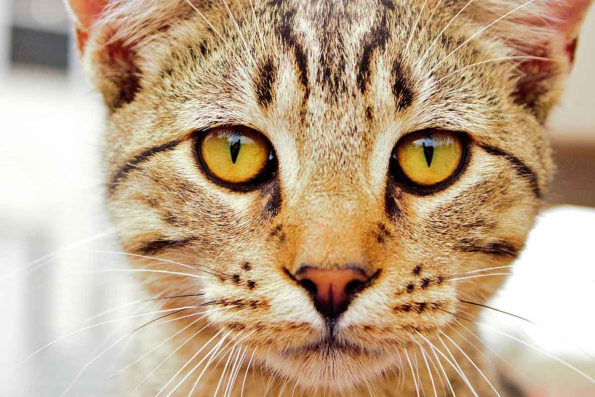
18. If you look carefully, you can see that the shape of the pupil in the sheep eye is oblong. A sheep's pupil is oblong, or rectangular, because it allows them to have a wider field of peripheral vision, which is crucial for prey animals like sheep to detect predators approaching from any direction, especially when grazing on the ground; the horizontal elongation of the pupil lets in more light from the sides, enhancing their ability to see around them effectively. Humans have round pupils because the muscles of the iris contract evenly, which helps us see small details in bright light. A cat's pupil is vertical because it helps them accurately judge the distance to their prey, a crucial ability for ambush predators like cats, as the vertical slit shape maximizes their depth perception by allowing for better stereopsis (comparing images from each eye to calculate distance) in low light conditions; essentially, the vertical slit provides a sharper vertical contour, aiding in precise distance calculations when hunting. See the pictures below:
 |
 |
 |
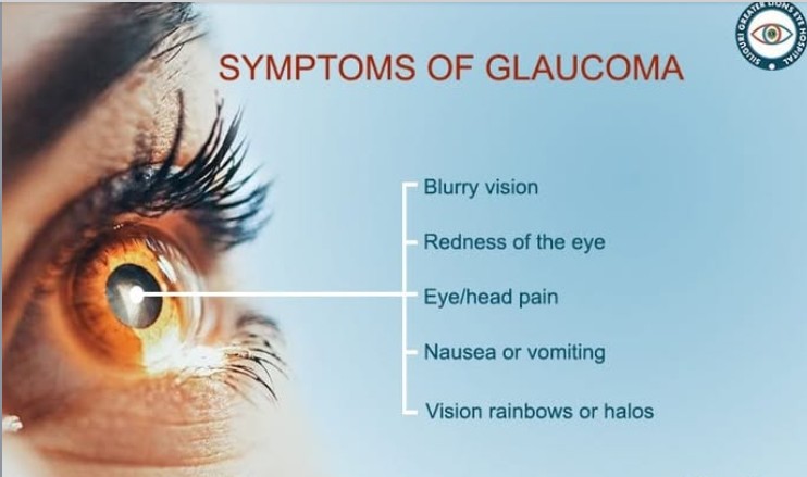
19. A second cavity can be found between the iris and the cornea. This layer is often perforated and empty, but it contained the watery liquid called the aqueous humor. This fluid, like the vitreous humor, helps maintain the shape of the eye. Glaucoma is a condition where the fluid pressure (called intraocular pressure) increases to the point where it puts pressure on the nerves of the retina, eventually causing them to die. Early symptoms of glaucoma include 1) gradual loss of peripheral (side) vision, 2) blurred or cloudy vision, and 3) difficulty seeing in dim light, 4) eye pain and discomfort. In severe cases nausea and vomiting can accompany these symptoms.
20. Remove the cornea from the front eye hemisphere. Use the scalpel to puncture a small slit at the boundary between the cornea and sclera. Then insert the scissors into the slit and cut all the way around the cornea to remove it. Notice the thickness of the cornea. Compared to the sclera, the cornea is slightly less thick than the sclera.
Cornea Thickness = 0.5 mm
Sclera Thickness = 1.0 mm
21. Discard the eye and clean all dissection utensils with warm water and soap. Dry them before putting them back into the kits. Wash and dry the blue pad in the dissection tray. Make sure that you lift the pad out and dry the underneath area as well.
Sheep Eye Dissection Pictures & Diagrams
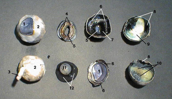
| 1. cornea 2. sclera 3. optic nerve 4. iris 5. pupil 6. ora serrata 7.ciliary body 8. choroid 9. tapetum lucidum 10. retina 11. lens 12. vitreous humor |
 |
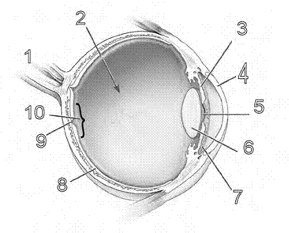
| 1. optic nerve 2. vitreous humor 3. iris 4. cornea 5. pupil 6. lens 7. aqueous humor 8.choroid 9. macula 10. retina |
 |
Vocabulary:
aqueous humor: a transparent water-like fluid similar to blood plasma, but containing low protein concentrations. It is secreted from the ciliary body, a structure supporting the lens of the eyeball.
ciliary body: a ring-shaped structure located in the eye, behind the iris and in front of the choroid. It plays a crucial role in maintaining the eye's shape and focusing light onto the retina.
cornea: the transparent, dome-shaped front layer of the eye that covers the iris and pupil; the cornea acts as a shield, protecting the eye from dirt, germs, and ultraviolet radiation.
suspensory ligaments: a series of fine, fibrous cords that connect the ciliary body to the lens of the eye. They play a crucial role in maintaining the position and shape of the lens, thus ensuring proper vision.
pupil: circular or oblong opening in the center of the iris (the colored part of the eye) that regulates the amount of light entering the eye.
glaucoma: a group of eye diseases that damage the optic nerve, which connects the eye to the brain. This damage is typically caused by increased pressure inside the eye (intraocular pressure).
macula: a small, oval-shaped area in the center of the retina, the light-sensitive tissue at the back of the eye. Its primary function is to provide sharp, central vision.
optic nerve: a sensory nerve that plays a crucial role in vision. The optic nerve is a vital sensory pathway that enables us to see and process visual information, contributing to our overall perception of the world around us. Damage to the optic nerve can result in vision loss or other visual impairment.
iris: the colored part of the eye that surrounds the pupil. Its primary function is to regulate the amount of light entering the eye.
sclera: commonly known as the "white of the eye," plays several crucial functions in maintaining eye health and vision. The sclera has a number of important functions: 1) structural support, 2) attachment for eye muscles, 3) protection from trauma, 4) blood vessel and nerve passage, 5) regulation of intraocular pressure, 6) vision enhancement, and 7) aesthetic function
ora serrata: a serrated junction in the eye that marks the transition from the retina to the ciliary body. It's an important anatomical landmark that helps separate the anterior and posterior chambers of the eye.
choroid: a vascular layer located between the retina and the sclera in the eye. It plays several crucial functions in maintaining eye health and vision: 1) nutrient supply, 2) heat regulation, 3) waste removal, 4) light absorption, 5) structural support, and 6) intraocular pressure regulation
tapetum lucidum: a reflective layer in the eye that enhances vision in low light conditions. It's found in many vertebrate animals including cats, dogs, sheep, cows, and owls.
retina: a light-sensitive layer of tissue located at the back of the eye. It plays a crucial role in vision by converting light into electrical signals that are sent to the brain.
lens: plays a crucial role in vision by focusing light onto the retina.
vitreous humor: also known as the vitreous gel, is a clear, jelly-like substance that fills most of the eye's volume. It performs several functions, including: 1) maintaining eye shape, 2) providing a clear pathway for light, 3) enabling oxygen and nutrient flow, 4) protecting the lens, and 5) playing a role in the development of the eye