|
Examine the photographs of the dorsal wall of the
body cavity of the male spiny dogfish shark by clicking the blue
lettered links in the column to the right. The specimen in the phographs
was prepared by removing almost the entire liver, alimentary canal,
pancreas, and spleen. This revealed the urogenital structures: gonads,
kidneys, and associated ducts.
The urinary and genital systems have distinct and
unique functions:
The function of the urinary system is to remove
nitrogenous wastes from the blood and maintain water balance.
The genital system is involved with the reproduction
of the species.
However, due to their similar developmental origins and the sharing of
common structures, they are usually considered as a single system.
The shark kidney and its ducts are quite different
from those in higher vertebrates. The relationship between the urinary
and genital structures is also quite different.
The kidneys are flattened, ribbon-like, darkly
colored structures Iying dorsally on either side of the midline, along
the entire length of the body cavity. A tough white glistening strip of
connective tissue is found between the kidneys in the midline.
The kidneys of the male are essentially the same as
those of the female. The posterior portion is involved in the
manufacture and transport of urine. The main difference lies in the
anterior portion of the kidney, which in females is degenerate and
functionless, but in males is an active part of the reproductive system.
|
Shark
Kidneys
Labeled
Kidneys
|
|
Examine the anterior view photographs of the shark by
clicking the blue lettered links in the column to the right.
Paired testes lie near the anterior end of the body
cavity, dorsal to the liver, adjacent to the anterior ends of the
kidneys.The sperm pass from the testes to the kidneys within narrow
tubules called efferent ductules.
|
Shark
Testes
Labeled
Testes
|
|
Examine the bottom view photographs of the shark by
clicking the blue lettered links in the column to the right.
After passing through the anterior end of the kidney
the sperm enter the ductus deferens and pass posteriorly toward the
cloaca. In mature male specimens the ductus deferens may be seen on the
ventral surface of the kidneys as a pair of highly coiled tubules.
Note: While in the female this duct carries urine, in
the male it transports spermatozoa and seminal fluid.
The posterior portion of the ductus deferens widens
and straightens to form the paired seminal vesicles.
|
Shark
Ductus Deferens
Labeled
Ductus Deferens
|
|
Examine the photographs of the shark's seminal
vesicles by clicking the blue lettered links in the column to the right.
The paired sperm sacs at the posterior ends of the
seminal vesicles receive the seminal secretions. They join to form the
urogenital sinuses which exit through the fleshy conical urogenital
papilla which extends from the cloaca.
The accessory urinary ducts, collect and transport
urine from the kidneys. These paired thin tubules may be found along the
medial side of the posterior half of the kidney. Small collecting
tubules from the kidneys lead into the accessory urinary ducts along
their lengths.
The cloaca receives the genital and urinary products
as well as the rectal wastes.
|
Shark
Seminal Vesicles
Labeled
Seminal Vesicles
|
|
Examine the photographs of the shark's claspers by
clicking the blue lettered links in the column to the right.
The claspers are modified extensions of the medial
portions of the pelvic fins. They are inserted into the female's cloaca
during copulation.
The finger-like claspers each have a dorsal groove,
the clasper tube that carries the seminal fluid from the cloaca of the
male to the cloaca of the female during mating.
|
Shark
Clasper Tubes
Labeled
Clasper Tubes
|
|
Examine the photographs of the dorsal wall of the
body cavity of the female spiny dogfish shark by clicking the blue
lettered links in the column to the right. The specimen in the phographs
was prepared by removing almost the entire liver, alimentary canal,
pancreas, and spleen. This revealed the urogenital structures: gonads,
kidneys, and associated ducts.
The ovaries are two cream-colored elongated organs in
the anterior part of the body cavity dorsal to the liver on either side
of the mid-dorsal line. The shape of the ovaries will vary depending
upon the maturity of the specimen. In immature females they will be
undifferentiated and glandular in appearance. In mature specimens you
may find two to three large eggs, about three centimeters in diameter,
in each ovary. When these break the surface of the ovary, upon
ovulation, they enter the body cavity and by means of peritoneal cilia
are moved into the oviducts.
The ovaries are the primary sex organs for the female
shark. They function to produce hormones to stimulate and maintain
sexual maturity.
|
Shark
Ovaries
Labeled
Ovaries
|
|
Examine the photographs of the female shark's
oviducts by clicking the blue lettered links in the column to the right.
The oviducts are elongated tube-like structures Iying
dorsolaterally the length of the body cavity, along the sides of the
kidneys. In mature specimens they are more prominent. The distal half of
the oviduct is enlarged to form the uterus.
The shell gland is the anterior end of the oviduct.
The eggs are fertilized and receive a light shell-like covering as they
pass through the shell gland.
The oviducts allow for the ova (singular = ovum) to
be transported from the peritoneal cavity.
|
Shark
Oviducts
Labeled
Oviducts
|
|
Examine the photographs of the female shark's uteri
by clicking the blue lettered links in the column to the right.
The posterior half of the oviduct becomes enlarged
and is known as the uterus. The fertilized eggs develop into embryos in
the uterus. Upon completing their period of gestation (close to two
years) the young are ready to be born.
The cloaca serves for the elimination of urinary and
fecal wastes as well as an aperture through which the young
"pups" are born.
The two uteri open into the posterodorsal portion of
the cloaca just ventral to the urinary papilla.
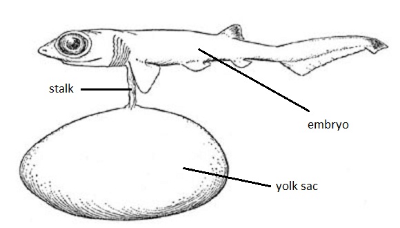
Fertilization in the dogfish shark is internal,
usually taking place within the shell gland of the oviduct. The
fertilized eggs continue to move posteriorly to the uterus. As they grow
the pups are attached to the egg, now known as the yolk sac, by means of
a stalk. During its period of gestation, which is nearly two years, the
yolk is slowly absorbed by the shark "pup."Numerous uterine
villi, finger-like projections from the uterine wall, make contact with
the surface ot the developing embryo and its yolk sac. It is believed
that these provide the embryo with water; all other nutrients are
supplied by the yolk. At birth the young are about 23 to 29 centimeters
long. This type of development, where the young are born as miniature
adults but have received hardly any nutrition directly from the mother's
uterus, is known as ovoviviparous.
|
Shark
Uteri
Labeled
Uteri
|
|
|
|
|
There are three types of embryonic
development: oviparous, ovoviviparous, and viviparous.
|
|
|
| 1. |
In oviparous ("egg
birth") sharks, a gland secretes a shell, or case,
around the egg as it passes through the oviduct,
protecting the shark until it hatches. The mother deposits
the egg cases in the sea.
|
| • |
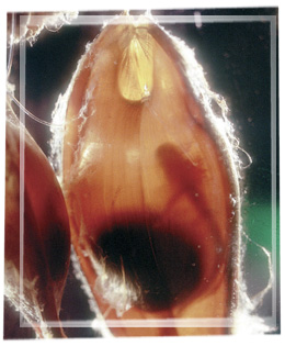
When the egg case is first laid,
it is soft and pale; the case hardens and darkens
in a few hours.
|
|
|
|
|
The egg
case, when it is first laid is soft and
pale.
|
|
|
| • |
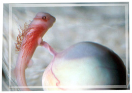
The developing embryo receives
nutrients from a yolk formed prior to
fertilization. |
|
|
|
|
|
A tiny shark embryo still
attached to its yolk.
|
|
|
|
| • |
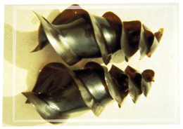
Oviparous sharks include horn
sharks and swell sharks (Cephaloscyllium
ventriosum). |
|
|
|
|
|
Horn sharks lay spiral
egg cases.
|
|
|
| • |
Port Jackson sharks carry their
egg cases in their mouths, possibly to drop them
in a hiding spot. This is about the only shark
parental care observed by humans. |
|
|
|
|
|
|
|
|
|
| 2. |
In ovoviviparous ("egg
live birth") sharks, the shell is often just a thin
membrane. Sometimes there is more than one egg in the
membrane; this group of eggs is called a candle. The
mother retains the egg, and the embryo soon sheds the
membrane and develops in the mother's uterus.
|
| • |
Theoretically, all the embryo's
nutrients come from the yolk. In some species,
however, the lining of the uterus probably
secretes nutritive fluids that are absorbed by the
embryo.
|
| • |
In other species,
embryos continue to obtain nutrients after their
yolk is absorbed by swallowing eggs and smaller
embryos in the uterus. This is termed
"intrauterine cannibalism" or ovophagy
("egg eating"). In these sharks, usually
only one embryo survives in each uterus. (Females
have two uteri). In simple terms....the
largest embryo will consume the smaller embryos,
so only one embryo will survive.
|
| • |
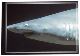
Ovoviviparous sharks
include mako sharks, dogfish sharks and sand tiger
sharks. |
|
|
|
|
|
Sand tiger sharks are
ovoviviparous.
|
|
|
|
|
|
| 3. |
In viviparous ("live
birth") sharks, the yolk stalk that connects the
embryo to the yolk grows long in the uterus. Where the
small yolk sac comes in contact with the mother's uterus,
it changes into a yolk sac placenta.
|
| • |
The embryo receives all its
nutrients from the mother in one of two ways:
|
| 1) |
Tissues of the embryo
and the mother are in intimate contact and
nutrients are passed directly from the
tissues of the mother to the tissues of
the developing embryo.
|
| 2) |
The uterine
lining secretes "uterine milk",
which bathes the developing embryo. The
branched yolk stalk absorbs the fluid.
|
|
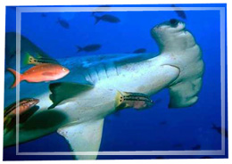
| • |
Viviparous sharks include
hammerhead sharks. |
|
|
|
|
|
Hammerhead sharks are
viviparous.
|
|
|
|
|
|
|
|
|
|
| 1. |
Gestation periods vary among species and
between individuals within a species. Since sharks and
batoids are ectothermic ("cold-blooded"), there
is no precise gestation time. The rate at which the embryo
develops depends on the water temperature. In general,
most embryos develop somewhere in the range of two months
(for some rays) to 18 to 24 months for the piked dogfish
(perhaps the longest of any vertebrate animal). Some
researchers believe basking sharks have a gestation period
of three and a half years.
|
|
|
|
