PART ONE:
Your Digestive System and How It Works
OVERVIEW OF THE DIGESTIVE SYSTEM:
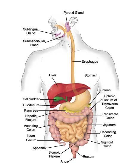
The digestive system is made up of the digestive tract—a series of hollow organs joined in a long, twisting tube from the mouth to the anus—and other organs that help the body break down and absorb food.
Organs that make up the digestive tract are the mouth, esophagus, stomach, small intestine, large intestine—also called the colon—rectum, and anus. Inside these hollow organs is a lining called the mucosa. In the mouth, stomach, and small intestine, the mucosa contains tiny glands that produce juices to help digest food. The digestive tract also contains a layer of smooth muscle that helps break down food and move it along the tract.
Two “solid” digestive organs, the liver and the pancreas, produce digestive juices that reach the intestine through small tubes called ducts. The gallbladder stores the liver’s digestive juices until they are needed in the intestine. Parts of the nervous and circulatory systems also play major roles in the digestive system.
You can see these organs in the diagram below
WHY IS DIGESTION IMPORTANT?
When you eat foods—such as bread, meat, and vegetables—they are not in a form that the body can use as nourishment. Food and drink must be changed into smaller molecules of nutrients before they can be absorbed into the blood and carried to cells throughout the body. Digestion is the process by which food and drink are broken down into their smallest parts so the body can use them to build and nourish cells and to provide energy.
HOW IS FOOD DIGESTED?
Digestion involves mixing food with digestive juices, moving it through the digestive tract, and breaking down large molecules of food into smaller molecules. Digestion begins in the mouth, when you chew and swallow, and is completed in the small intestine.
MOVEMENT OF FOOD THROUGH THE DIGESTIVE SYSTEM
The large, hollow organs of the digestive tract contain a layer of muscle that enables their walls to move. The movement of organ walls can propel food and liquid through the system and also can mix the contents within each organ. Food moves from one organ to the next through muscle action called peristalsis. Peristalsis looks like an ocean wave traveling through the muscle. The muscle of the organ contracts to create a narrowing and then propels the narrowed portion slowly down the length of the organ. These waves of narrowing push the food and fluid in front of them through each hollow organ.
The first major muscle movement occurs when food or liquid is swallowed. Although you are able to start swallowing by choice, once the swallow begins, it becomes involuntary and proceeds under the control of the nerves.
See the diagrams below for a better understanding of peristalsis.
|
|
|
Swallowed food is pushed into the esophagus, which connects the throat above with the stomach below. At the junction of the esophagus and stomach, there is a ringlike muscle, called the lower esophageal sphincter (sometimes also called the cardiac sphincter), closing the passage between the two organs. As food approaches the closed sphincter, the sphincter relaxes and allows the food to pass through to the stomach.
See the diagrams below for a better understanding of the esophagus and the lower esophageal or cardiac sphincter.
|
|
|
The stomach has three mechanical tasks. First, it stores the swallowed food and liquid. To do this, the muscle of the upper part of the stomach relaxes to accept large volumes of swallowed material. The second job is to mix up the food, liquid, and digestive juice produced by the stomach. The lower part of the stomach mixes these materials by its muscle action. The third task of the stomach is to empty its contents slowly into the small intestine.
See the diagrams below of the stomach and its lining. Notice the layers of muscle and how they are orientated at right angles to each other to add strength to the muscle.
|
|
 |
There are several "anatomical" divisions to the stomach. The fundus is found to the left and upward from the esophageal opening to the stomach. The body is the middle area of the stomach and the pylorus is the lower area of the stomach. As the stomach sits in place in the body cavity you can see that it has two curves. The "outer" curve is known as the greater curvature (larger), whereas the "inner" curve is called the lesser curvature (smaller).
See the diagram below for more detail of the stomach.
|
|
Several factors affect emptying of the stomach, including the kind of food and the degree of muscle action of the emptying stomach and the small intestine. Carbohydrates, for example, spend the least amount of time in the stomach, while protein stays in the stomach longer, and fats the longest. As the food dissolves into the juices from the pancreas, liver, and intestine, the contents of the intestine are mixed and pushed forward to allow further digestion.
The small intestine in an adult human measures on average about 5 meters (16 feet), with a normal range of 3 - 7 meters; it can measure around 50% longer at autopsy because of loss of smooth muscle tone after death. It is approximately 2.5-3 cm in diameter. Although the small intestine is much longer than the large intestine (typically around 3 times longer), it gets its name from its comparatively smaller diameter. Although as a simple tube the length and diameter of the small intestine would have a surface area of only about 0.5m2, the surface complexity of the inner lining of the small intestine increase its surface area by a factor of 500 to approximately 200m2, or roughly the size of a tennis court.
The small intestine is divided into three structural parts: the duodenum, the jejunum, and the ileum.
The duodenum
is the first section of the small intestine and is the shortest part of the
small intestine. Most chemical digestion takes place here and it is the
principle site for iron absorption. The name duodenum is from the Latin duodenum
digitorum or "twelve fingers' breadths.
The jejunum
is the middle section of the small intestine.
The ileum
is the final section of the small intestine. The ileum follows the
duodenum and jejunum and is separated from the cecum by the ileocecal valve (ICV).
In humans, the ileum is about 2-4 m long, and the pH is usually between 7 and 8
(neutral or slightly alkaline).
See the diagrams below of the small intestine and its lining to help you understand how it absorbs the nutrients.
|
|
 |
|
|
|
 |
 |
What ever is not absorbed by the small intestine will enter the large intestine.
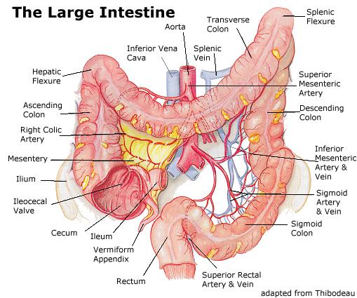
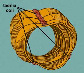
The large intestine extends from the ileocecal junction to the anus and is about 1.5m long. On the surface, you can identify bands of longitudinal muscle fibers called taeniae coli, each about 5mm wide. There are three bands and they start at the base of the appendix and extend from the cecum to the rectum. The function of the bands is to add strenght and stability to the large intestine. Along the sides of the taeniae, you will find tags of peritoneum filled with fat, called epiploic appendages (or appendices epiploicae). The sacculations, called haustra, are characteristic features of the large intestine, and distinguish it from the rest of the intestinal tract.
See the diagrams below of the large intestine and the taeniae coli.
|
|
 |
The cecum is about 6cm long and is a blind cul-de-sac
which lies in the right iliac fossa. It is the part of the colon below the
opening of the ileum into the colon. The cecum lies immediately behind the
abdominal wall and greater omentum. There is frequently a peritoneal recess
behind the cecum called the retrocecal recess and
the appendix is sometimes hiding within this recess and may extend as far
superiorly as the liver.
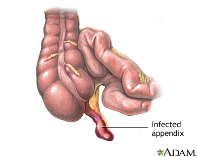
Hanging off the cecum is the vermiform appendix
which opens into the cecum about 2cm below the ileocecal opening. The average
length of the appendix is about 10cm and may lie in different positions. It has
its own mesentery called the mesoappendix which
carries the appendicular artery.
See the diagrams below for a better idea of the location of the appendix.
|
|
 |
The functions of the large intestine include:
Mechanical digestion. Rhythmic contractions of the large intestine produce a form of segmentation called haustral contractions in which food residues are mixed and forced to move from one haustrum to the next. Peristaltic contractions produce mass movements of larger amounts of material.
Chemical digestion. Digestion occurs as a result of bacteria that colonize the large intestine. They break down indigestible material by fermentation, releasing various gases. Vitamin K and certain B vitamins are also produced by bacterial activity.
Absorption. Vitamins B and K, some electrolytes (Na+ and Cl−), and most of the remaining water is absorbed by the large intestine.
Defecation. Mass movement of feces into the rectum stimulates a defecation reflex that opens the internal anal sphincter. Unless the external and sphincter is voluntarily closed, feces are evacuated through the anus. Human feces (also faeces), also known as stools, is the waste product of the human digestive system and varies significantly in appearance, depending on the state of the whole digestive system, influenced and found by diet and health. Normally stools are semisolid, with a mucus coating. Small pieces of harder, less moist feces can sometimes be seen impacted on the distal (leading) end. This is a normal occurrence when a prior bowel movement is incomplete; and feces are returned from the rectum to the intestine, where water is absorbed.
There are several other organs that are involved in digestion as well. These "solid" organs do not have food traveling through them, but they are just important and they are the liver and the pancreas.
The liver is a vital part of the digestive system. It has a wide range of functions, including detoxification, protein synthesis, and production of biochemicals necessary for digestion. The liver is necessary for survival; there is currently no way to compensate for the absence of liver function.
This organ plays a major role in metabolism and has a number of functions in the body, including glycogen storage, decomposition of red blood cells, plasma protein synthesis, hormone production, and detoxification. It lies below the diaphragm in the thoracic region of the abdomen. It produces bile, an alkaline compound which aids in digestion, via the emulsification of lipids, which basically means it helps to break down fats into a usable form for the cells. It also performs and regulates a wide variety of high-volume biochemical reactions requiring highly specialized tissues, including the synthesis and breakdown of small and complex molecules, many of which are necessary for normal vital functions.
See the diagrams of the liver below for more understanding.
 |
 |
The other organ that is a vital part of the digestive system is the pancreas.
The pancreas is a gland organ in the digestive and endocrine system of humans. It is both an endocrine gland producing several important hormones, including insulin, glucagon, and somatostatin, as well as an exocrine gland, secreting pancreatic juice containing digestive enzymes that pass to the small intestine. These enzymes help in the further breakdown of the carbohydrates, protein, and fat in the chyme.
See the diagrams below to learn more about the pancreas.
 |
 |