
 |
 |
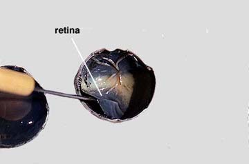
| Step 5: Arrange the two hemispheres of the eye as you see in the left photograph. Observe the semi-fluid vitreous humor that fills the central cavity of the eye. It is transparent in the living eye but might be cloudy in the preserved specimen. The vitreous humor along with the aqueous humor helps to maintain the shape of the eye. More will be said about the aqueous humor later. The retina lines the the posterior cavity of the eye and extends forward to the ciliary body. Use your probe to lift and pull the retina back from the underlying choroid layer. See the photograph on the right side above. Notice that the retina is only firmly attached to the choroid at one place. This region is the optic disc or blind spot. Here the nerve fibers leave the retina and form the optic nerve which is directly behind the blind spot. Take the notes you need to record what you have observed so far. | |